Biomedical Engineering Reference
In-Depth Information
16
(A)
(B)
14
12
10
8
6
4
2
0
500 nm
Nano apatite crystal length (nm)
(C)
O
O
50
µ
m
O
O
O
(E)
(D)
500 nm
P
E
Apatite
nanocrystals
P
C
100µm
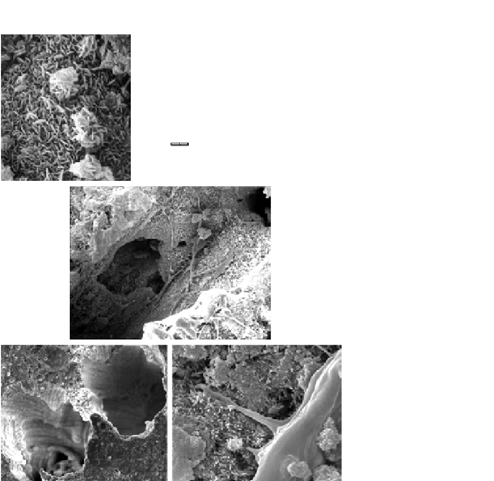
FIGURE 12.1
CPC-based nano-apatite scaffold. (A) SEM of the nano-sized hydroxyapatite crystals
that make up CPC. (B) Size distribution of apatite crystals in CPC. The crystal length
ranged from 75 to 550 nm. The width ranged from 50 to 150 nm. (C) Macroporosity
was created in CPC via the dissolution of mannitol porogen, and pre-osteoblastic cells
(mouse MC3T3-E1 cells) (indicated by “O”) in the pore. (D) Macropore channels “P”
in CPC formed via fibers after fiber dissolution. (E) Human umbilical cord stem cells
(hUCMSC) attaching to the nano-apatite in CPC via cytoplasmic extensions. “C” refers
to the hUCMSC. “E” designates the cytoplasmic extensions of the cell.