Biomedical Engineering Reference
In-Depth Information
(A)
(B)
(C)
(D)
(E)
PLLA/HA
PLLA/MWCNTs/HA
Calcium Ka1
Calcium Ka1
1800
2000
1000
1200
1400 1600
Raman shift (cm
-1
)
Phoshphorous Ka1
Phoshphorous Ka1
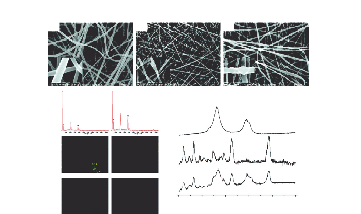
FIGURE 10.4
Characteristics of three kinds of membranes: (A-C) represent typical scanning electron microscope
(SEM) images of PLLA, PLLA/HA, and PLLA/MWCNTs/HA membranes, respectively; (D) represents the
EDX mapping of PLLA/HA (left) and PLLA/MWCNTs/HA (right) membranes for Ca (middle) and P (lower)
elements; (E) represents Raman spectra of MWCNTs (upper), PLLA/HA membrane (middle), and PLLA/
MWCNTs/HA membranes (lower).
10.4.4
Cell Culture on PLLA/MWCNTs/HA Composite Nanofibers Membranes
PDLCs were obtained from healthy teeth extracted for orthodontic reasons. GECs were obtained
from the gingival tissue of systemic healthy individuals removed during periodontal surgery. Both
the cells between the third and the fifth passages were used in the following studies. PDLCs and
GECs were harvested with 0.25% trypsin/0.02% Ethylenediaminetetraacetic acid (EDTA), and PDLCs
were transferred to an osteogenic differentiation medium, α-MEM containing 10% fetal bovine serum
and antibiotics supplemented with 10 nM dexamethasone, 10 mM β-glycero-phosphate, and 50 mg l
1
of ascorbic acid. PDLCs or GECs were seeded onto PLLA, PLLA/HA, and PLLA/MWCNTs/HA
fibrous membranes, as well as tissue culture polystyrene (TCPS; control group) at a density of 5,000
cells/well (24-well culture plate).