Biomedical Engineering Reference
In-Depth Information
advance in microscope design since the construction of compound mi-
croscopes. The transmission electron microscope allowed scientists not
only to look at the surface, but to peer inside, living cells and discover
their complexity. It allowed us to see viruses for the very first time, and
it also made it possible to look at cell membranes, and even some large
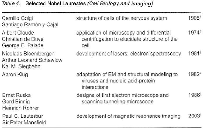
protein complexes. Ernst Ruska was award the Nobel Prize in 1986 for
the development of the transmission electron microscope, a prize he
shared with two scientists, Heinrich Rohrer and Gerd Binnig, for their
development of the scanning tunneling microscope (Table 4), which is
used to study atoms on physical surfaces, such as silicon chips for
computers. However, because samples that are being visualized in an
electron microscope must be maintained in a vacuum, this technology
does not allow cell biologist to study details of living cells.
The advent of fluorescent microscopic techniques by Albert H. Coons,
using “labeled” antibodies and other probes to directly visualize tissues
and components cells, and the coupling of the microscope to high res-
olution digital cameras and the computer, has truly revolutionized the
use of microscope. Not only does direct transfer of images from a digital
still or movie camera into a microscope allow an investigator to capture
and store data, but it has promoted the development of sophisticated
imaging software as well. Such software programs allow images to be
significantly enhanced, extending the resolution of the light microscope.
It is now possible to look within live cells and document the movements

Search WWH ::

Custom Search