Biomedical Engineering Reference
In-Depth Information
(A)
(B)
2mm
(D)
(C)
100
µ
m
50
µ
m
FIGURE 3.6
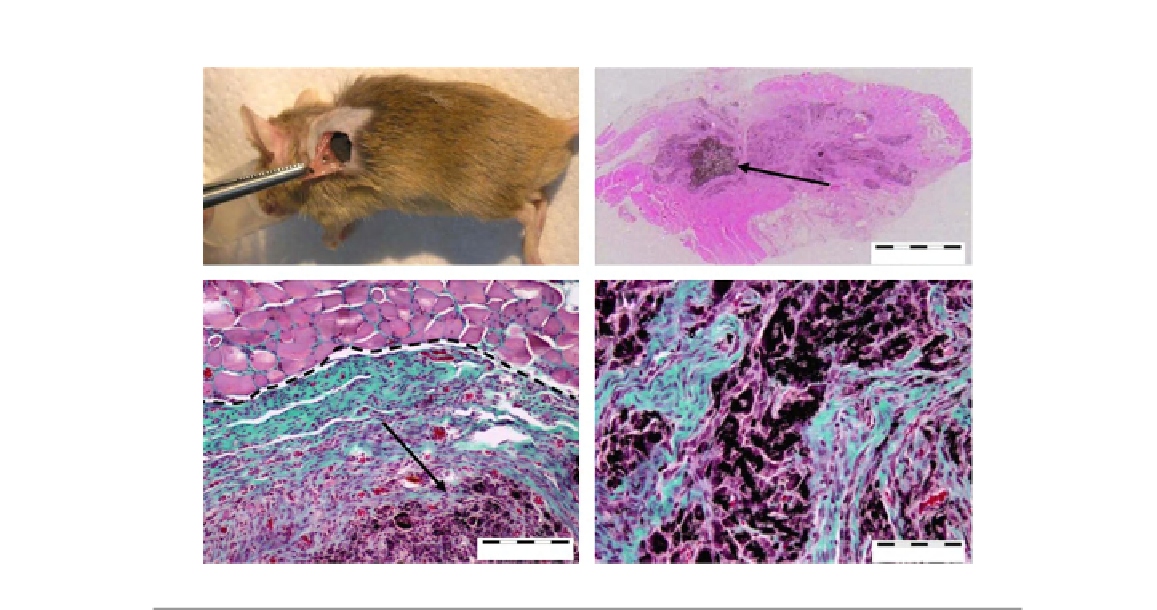
Picture (A) shows the surgery implantation of rhBMP-2 adsorbed MWNT/CHI (chitosan) scaffolds into mouse
subcutaneous muscular pocket. Optical microscope micrograph (B) shows regenerated bone tissue and a
minor fraction of remaining MWNT/CHI scaffold. Optical micrograph (C) shows a detail of regenerated bone
tissue (collagen expressing cells, bluegreen colored) after major disassembly of the MWNT/CHI scaffold,
surrounded by muscle tissue (pink colored). The well-limited interface between adjacent tissues is
remarkable (see black dash line). The remaining MWNT/CHI scaffold (black colored) is pointed by black
arrow. Optical micrograph (D) shows a detail of remaining scaffold plenty of fibroblasts (purple colored), prior
to its disassembly and colonization by collagen expressing cells (blue
green colored). (For interpretation of
the references to color in this figure legend, the reader is referred to the web version of this topic.)
From Ref.
[248]
.
Due to its nanoscale size, solubility, reduced cellular toxicity, and cationic surface of functiona-
lized CNT, its potential use as a novel drug delivery vehicle has been studied extensively.
Functionalized CNT conjugated with peptides was able to penetrate into the cells such as HeLa
immortal cells, fibroblasts, and keratocytes, indicating the potential of functionalized CNT as a
carrier for drug delivery
[253]
.
A group of researchers conjugated SWNT with the anticancer drug cisplatin and a receptor
ligand, epidermal growth factor (EGF) to treat squamous cell carcinoma, which is one of the most
common oral cancers
[254]
. By having EGF, which has a strong affinity to the cell-surface receptor
which is overexpressed in squamous cell carcinoma, the compound was able to target cancer cells
with high specificity. Authors found that functionalized and bioconjugated CNT caused endocytosis
by drawing CNT into the cell.