Biomedical Engineering Reference
In-Depth Information
FIGURE 21.3
SEM image revealing the dentin
nanocomposite interface. The dentinal tubules (right) are seen to be
penetrated by the nanocomposite fibers.
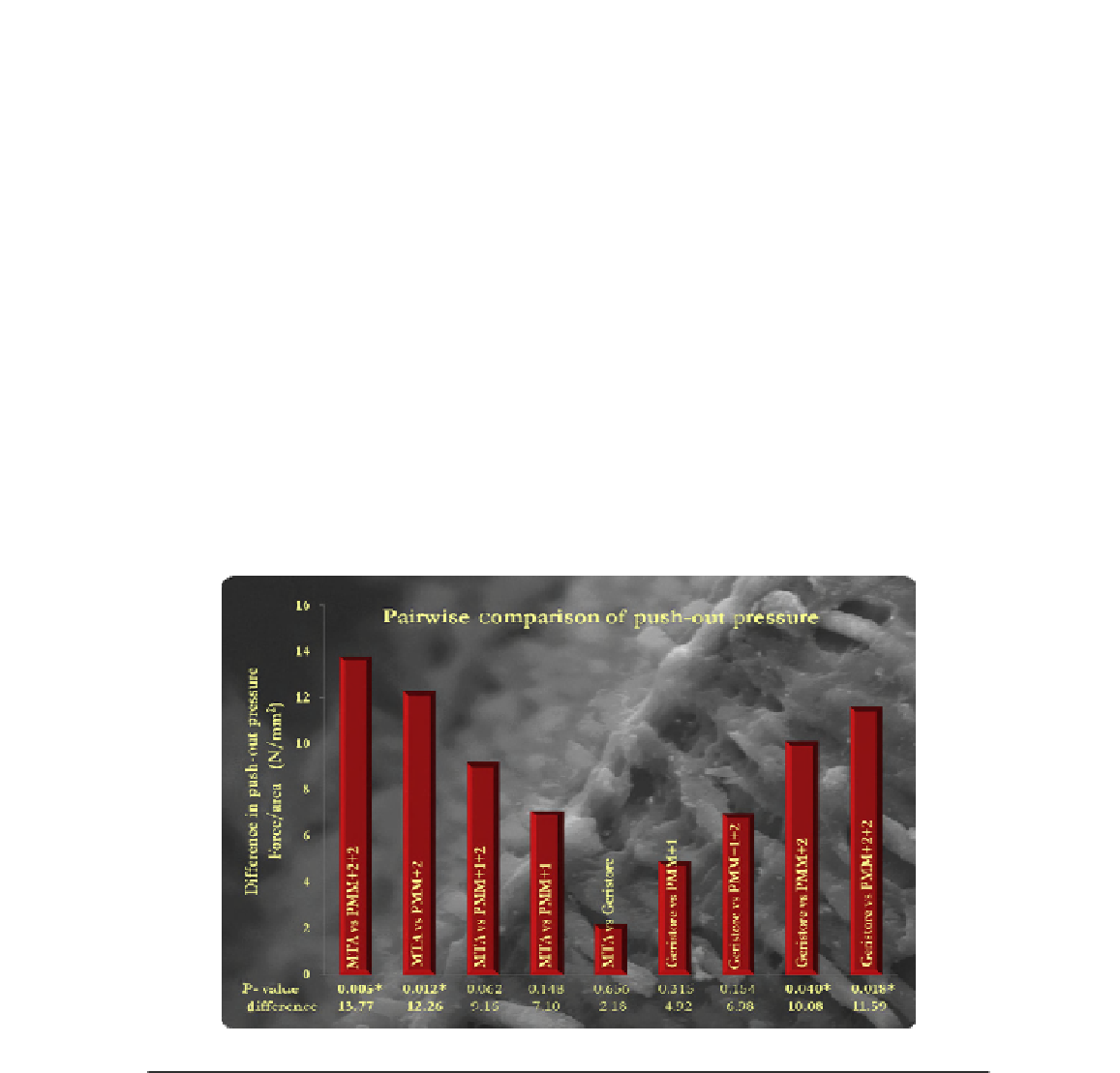
FIGURE 21.4
Bar chart comparing push-out force for nanocomposites Poly(methyl methacrylate) (PMM) containing varying
quantities of organoclay nanoparticles (1
2%) with MTA and Geristore. The 2% nanocomposite groups
performed significantly better than MTA and Geristore.