Biomedical Engineering Reference
In-Depth Information
reinforce PPF to cover the limitations of inferior mechanical properties of PPF for use in load-
bearing applications. On the other hand, many reports have revealed that the incorporation of CNTs
into polymeric matrix could render nanocomposite scaffolds with some osteogenic and bioactive
properties. Pan et al.
[24]
found that the scaffolds with low concentration (0.5 wt %) of MWCNTs
were able to enhance the proliferation and differentiation of rat BMSCs. Sitharaman et al.
[26]
evaluated the in vivo biocompatibility of US-tube-reinforced PPF scaffolds in a rabbit model. US-
tube nanocomposite scaffolds and control polymer scaffolds were implanted in rabbit femoral con-
dyles and in subcutaneous pockets. At 4 and 12 weeks after implantation, examinations showed
that the porous US-tube nanocomposite scaffolds exhibited favorable hard and soft tissue responses
at both time points. At 12 weeks, US-tube nanocomposite scaffolds had promoted a three-fold
greater bone tissue ingrowth than control polymer scaffolds. As shown in
Figure 18.3
, both PPF
(A)
(B)
100
μ
m
100
μ
m
(C)
(D)
100
μ
m
100
μ
m
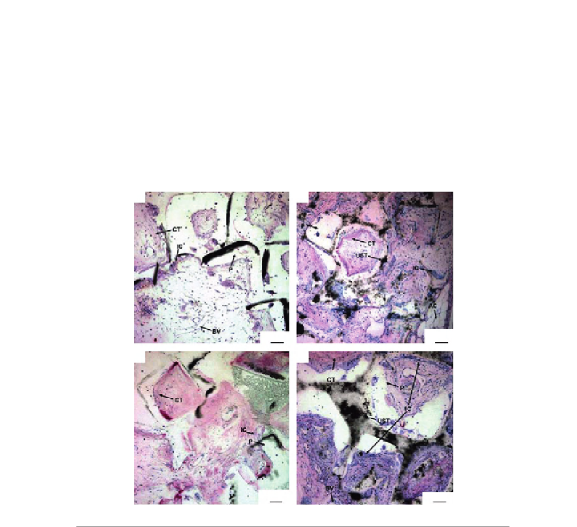
FIGURE 18.3
Representative histological sections of scaffolds implanted subcutaneously: (A) a PPF scaffold 4 weeks after
implantation, (B) a US-tube/PPF scaffold after 4 weeks, (C) a PPF implant after 12 weeks, and (D)
a US-tube/PPF implant after 12 weeks. The images are presented at 10
magnification. P: PPF scaffold,
UST: US-tubes, CT: connective tissue, AC: adipose cells, BV: blood vessels, IC: inflammatory cells.
Reproduced with permission from Ref.
[26]
.
3