Biomedical Engineering Reference
In-Depth Information
Bacteria
Receptor
Adhesion
Glucans
Enamel
Pellicle
Dentin
Enamel
Pulp
(A)
(B)
ACP
CPP
II
III
Bacteria
Shear forces
in the mouth
Ca
2+
I
II
Nanocomposite
Pellicle
Enamel
(C)
(D)
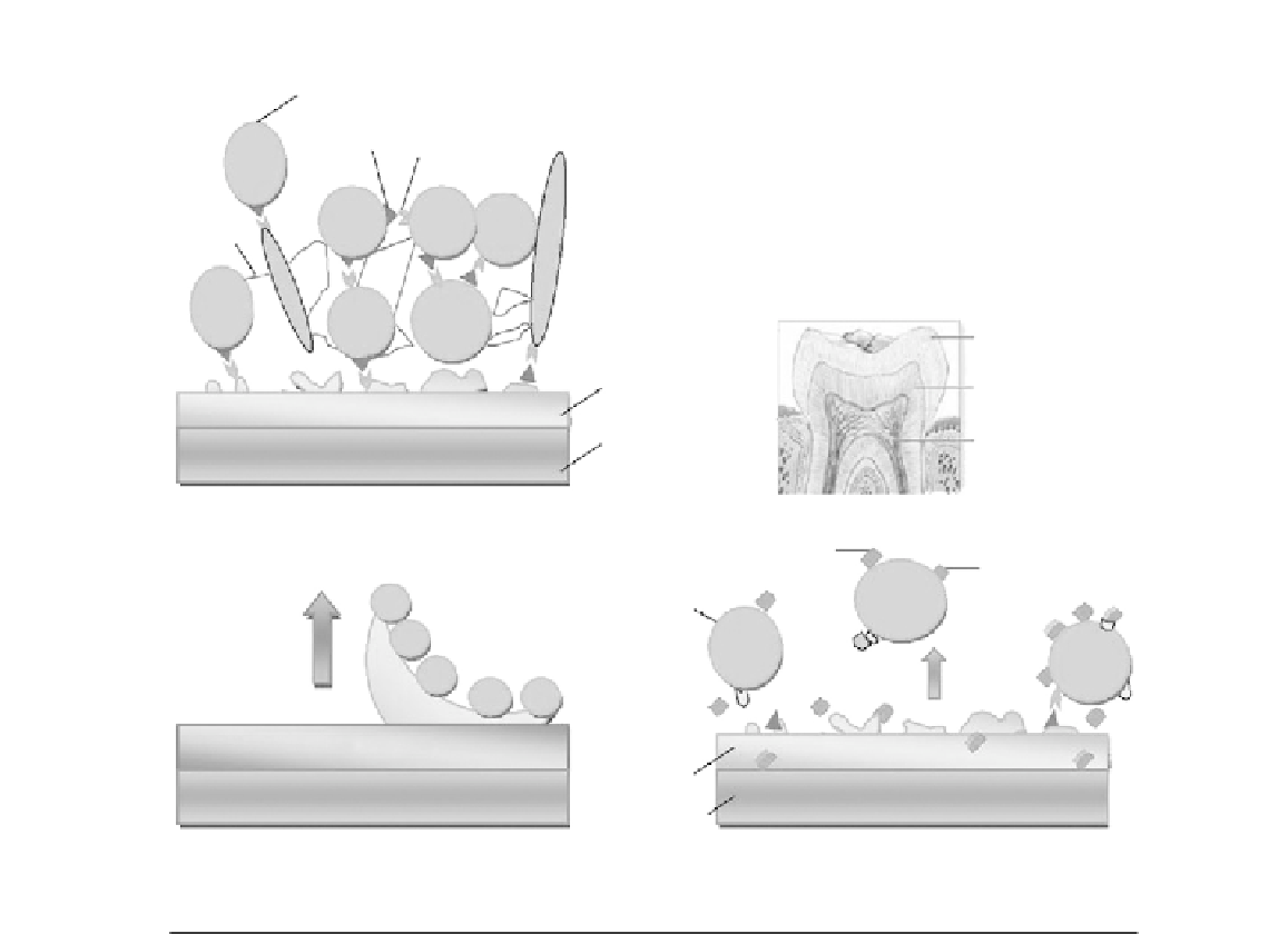
FIGURE 2.1
Bioadhesion and biofilm management in the oral cavity. (A) Bioadhesion in the oral cavity. Proteins interact
with the enamel surface to form a proteinaceous pellicle layer. Bacteria adhere to this conditioning film
through calcium bridges and specific adhesion
receptor interactions. Bacteria are surrounded by an
extracellular matrix of water insoluble glucans, and they communicate through quorum sensing (arrows). (B)
Cross section of a human molar tooth showing the enamel, dentin, and pulp chamber. (C) Easy-to-clean
nanocomposite surface coating. The low-surface-free-energy coating (circles) causes poor protein
protein
binding. Shear forces in the mouth can easily detach the outer layer of the pellicle and bacterial biofilm from
the surface. (D) CPP
ACP inhibits bacterial adhesion and oral biofilm formation. CPP attaches to the pellicle
and limits bacterial adhesion. It competes with calcium for plaque
calcium binding sites (I), and decreases
the amount of calcium bridging the pellicle and bacteria, and between the bacterial cells. Specific receptor
molecules in the pellicle layer and on the bacterial surfaces are blocked; further reducing adhesion and
coadhesion (II). This affects the viability of the bacteria (III)
[36]
.