Biomedical Engineering Reference
In-Depth Information
7.3.1
Etch-and-rinse adhesives
These adhesives utilize the use of acid etching of both enamel and dentin and the application of
both primer and adhesive in separate steps or the application of the primer/adhesive mixture in a
single bonding step. Phosphoric acid with a concentration of 30
40% is the acid of choice for such
adhesive systems. Although enamel etching was established since Buonocore's study, acid etching
of dentin was not recognized until Fusayama
[8]
introduced the total-etch concept. One of the
causes for previously reported failures of such concept was the dry bonding technique, until Kanca
[9]
described the so-called “wet bonding” (
Figure 7.4
). In this regard, the complete demineraliza-
tion of dentin with phosphoric acid etching gel is limited to the superficial 5
m(
Figure 7.5
)of
intertubular dentin
[10]
, which is followed by the application of solvated primer or solvated primer/
adhesive mixture to gain retention through the formation of microresin tags.
Ideally, the resin monomer should completely replace the lost minerals (
Figure 7.5
). This could
never be ideally performed
[9]
, which is due to the presence of residual solvent or due to the fluid
infiltration through the movement of dentinal fluid from inside the dentinal tubules
[11,12]
. This
will cause incomplete infiltration of monomer inside the created porosities (
Figures 7.6 and 7.7
).
Incomplete infiltration of resin monomer is one of the causes that make the etch-and-rinse adhe-
sives technique sensitive.
8
µ
7.3.2
Self-etching adhesives
Self-etching adhesives can be classified into four categories according to the pH of the functional
monomer: the strong self-etching adhesives (pH
,
1), intermediate adhesives (pH
1.5), mild adhe-
sives (pH
2)
[13]
, and extra-mild adhesives (pH
.
2.5)
[14]
.
2
µ
m
500 nm
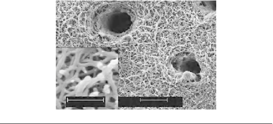
FIGURE 7.4
Scanning electron micrograph of acid-etched dentin showing two dentinal tubules containing remnants of
peritubular dentin matrix. Inset: High magnification of branching collagen fibrils (ca. 75 nm in diameter)
separated by interfibrillar spaces that serve as channels for resin infiltrations during bonding.