Biomedical Engineering Reference
In-Depth Information
It is speculated that the nanofiller components also enhance some physical properties of the
hardened restorative. However, in a study it is reported that the KN showed low hardness value
(39 KHN) when compared with other resin-modified glass ionomer, Vitremer (69.9 KHN). It is
suggested that this nanofilled GIC could be indicated to anterior teeth or cervical restorations and
does not seem to be appropriate to use in stress-bearing areas. However, according to the manufac-
turer (3M ESPE), KN is indicated for small Class I restorations, Class II and V, sandwich
technique, primary teeth restorations, and provisional restorations. It is wise to observe that the
material does not comply with the specifications of ADA (American Dental Association), which
regulates the number of Knoop hardness of ionomer material indicated for restoration in 48 KHN
[46]
. Whereas, recently in another study, El Halim
[47]
found higher values of microhardness
(62 KHN) of KN, which are in agreement of ADA specifications.
Bond strength of nanoionomer was analyzed and it was observed that the material interacted with
dentin and enamel in a very superficial way, without evidence of demineralization and/or hybridization.
They exhibited adequate bond strength to enamel (14.4
6
5.8 MPa) and dentin (12.6
6
6.5 MPa), at the
same extent as other GICs (enamel: 12.9
2.9 MPa), on the condition that
the surface was beforehand treated with the proprietary primer that contains the acrylic/itaconic acid
copolymer dissolved in HEMA and water. It bonded less effectively than conventional RMGIC (Fuji II
LC) (enamel (38.8
6
3.3 MPa; dentin: 12.3
6
4.3 MPa)). Its superficial interaction provides micro-
mechanical interlocking that is most likely supported by chemical interaction of the acrylic/itaconic
acid copolymer with surface HA
[48]
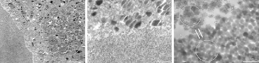
. The transmission electron microscope (TEM) images shown in
Figure 5.1
exhibited the nonhomogeneous filler distribution and there are some nanoclusters.
Another study using the shear bond strength (SBS) as an adhesion parameter showed that Er:YAG
laser dentin pretreatment results in lower bond strength values compared to acid-etching or a combined
acid-etching and laser pretreatment
[49]
. A study
[50]
on bonding orthodontic brackets showed
6
7.4 MPa) and dentin (31.4
6
(A)
(B)
(C)
FIGURE 5.1
TEM images of the nano-RMGIC, disclosing areas of nonhomogeneous filler distribution at certain locations
(A). An area of highly packed mixed glass particles is shown (round frame), as well as a cluster of nanofillers
clearly apart (arrowheads) from the remaining microstructure. The rectangular frame delimits an area
examined in higher magnification (B), which shows the interface between a highly packed cluster of nanofiller
and the remaining microstructure (arrowheads). The rectangular frame delimits an area examined at higher
magnification (C), which displays a detail of the interface (arrowheads) between the nanofiller cluster and the
nanofillers in the typical microstructure
[48]
.