Biomedical Engineering Reference
In-Depth Information
has a unique structure with a triple helical conformation that allows
N,N
-dimethyl-2,7-diazapyrenium dication (DMDAP) and PDGF
bindings. The luorescence of DMDAP is almost completely quenched
by Apt-Au NPs when it intercalates with the aptamers. Once PDGFs
bind to the Apt-Au NPs, the DMDAP molecules are released from the
NP surfaces into the bulk solution, leading to increased luorescence.
Owing to the large magnitudes of these increases (up to 40-fold) in
the turn-on luorescence signals of DMDAP/Apt-Au NPs upon PDGFs
binding, this approach is highly sensitive for the detection of PDGFs.
(A)
(B)
(C)
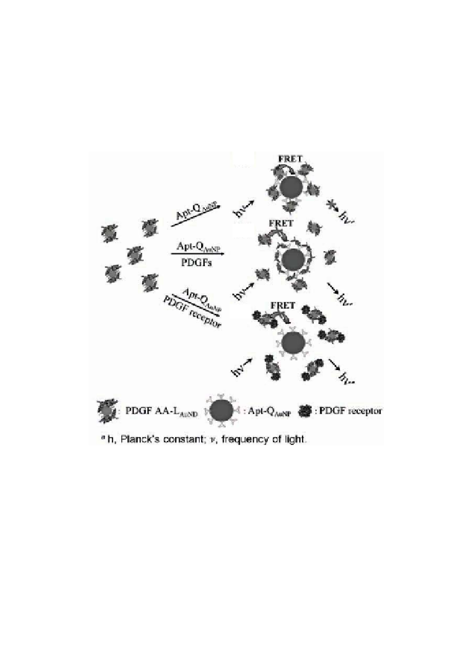
Figure 10.8
Schematic representations of PDGF and PDGF receptor
nanosensors that operate based on the modulation of the
photoluminescence quenching between PDGF AA-L
AuND
and
Apt-Q
AuNP
. Reprinted with permission from Ref. 86. See also
Color Insert.
Use of two differently sized Au NPs, acting separately as donors
and acceptors, in homogeneous photoluminescence quenching
assays are sensitive for the analysis of proteins.
86
Introduction
Search WWH ::

Custom Search