Biomedical Engineering Reference
In-Depth Information
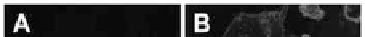
a nuclear dye, SYTO 59, allowed observation of the intracellular and
nuclear distributions of the NLS-11-MUA-Au NCs inside the cells.
The two-color co-localization images (panels D and F) reveal that the
blue luminescence from the NLS-11-MUA-Au NCs was distributed
well within both the cytoplasm (panel D) and the nucleus (panel
F); in contrast, the image of the cells treated with the 11-MUA-Au
NCs (panel B) exhibits only the red luorescence of WGA-Alexa 594.
Therefore, the nuclear targeting of 11-MUA-Au NCs was possible
because of the functionality of NLS, as revealed in panel F with the
two-color co-localization images in the same confocal z-plane (blue:
NLS-11-MUA-Au NCs; red: SYTO 59).
Figure 9.8
Confocal microscopy images of intracellular delivery of the Au
NCs. HeLa cells were treated with 11-MUA-Au NCs (A, B) and
NLS−11-MUA−Au NCs (E, F) for 1.5 h. The left panels present
one-color images; the right panels present two-color co-
localization images of the HeLa cells incubated with NLS-11-
MUA-Au NCs and counterstained with a speciic membrane
dye (WGA-Alexa 594) and a nuclear dye (SYTO 59). Scale bar:
25 μm. Reprinted with permission from Ref. 72. See also Color
Insert.
Water-soluble, luorescent, thiol-capped Au NPs can also be
selective and sensitive probe for the detection of various analytes
of interest, such as heavy metal ion pollutants, which exert adverse
effects on the environment and on human health. 11-MUA-Au


Search WWH ::

Custom Search