Biomedical Engineering Reference
In-Depth Information
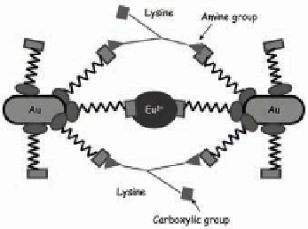
Figure 8.6
Schematic for the MUA-Au-NP resonance light scattering plas-
mon enhancement model. The RLS enhancement is achieved
through cooperative binding with europium and lysine.
Reprinted from Ref. 37 with permission.
forces among the negatively charged MUA-Au-NPs are strong
enough to hinder the electrostatic attraction between the positively
charged amino groups in lysine and the negatively charged MUA-
Au-NPs. Because the RLS intensity of Eu
3+
ion decreases upon
increasing MUA-Au-NPs concentration, energy transfer from Eu
3+
ions to MUA-Au-NPs occurs, signiicantly enhancing the eficiency of
interplasmon coupling. In other words, Eu
3+
ions act as sensitizers
for the RLS emission (390 nm) of the Au NPs. Figure 8.7 displays
1000
1000
800
800
600
600
400
400
200
200
0
300
0
300
350
350
400
400
450
450
500
500
550
550
600
600
650
650
λ (nm)
Figure 8.7
RLS spectra of: (a) MUA-Au-NPs; (b) MUA-Au-NPs-Lysine; (c)
MUA-Au-NPs-Eu 1 × 10
-3
M-Lysine; (d) MUA-Au-NPs-Eu 2.5 ×
10
-3
M-Lys; (e) MUA-Au-NPs-Eu 3.0 × 10
-3
M-Lysine; (f) MUA-
Au-NPs-Eu 5.0 × 10
-3
M-Lysine; (g) MUA-Au-NPs-Eu 7.5 × 10
-3
M-Lysine. Inset: Confocal luorescence microscopy images of
(a) agglomerated locs of MUA-Au-NPs-Eu-Lysine and (b)
isolated clusters of MUA-Au-NPs-Eu-Glutamine. [MUA-Au-NPs]
= 5.3 × 10
-12
M, [L-lysine] = 7.0 × 10
-3
M, [Glutamine] = 7 ×
10
-3
M, pH = 6. Reprinted from Ref. 37 with permission. See
also Color Insert.
λ (nm)


Search WWH ::

Custom Search