Biomedical Engineering Reference
In-Depth Information
eukaryotic. algal. organisms. that. comprise. only. one. cell,. are. photosynthetic,. and. have. a.
long.scientiic.history.of.study,
43,44
.which.is.partly.due.to.the.astonishingly.intricate.design.
and.architecture.of.their.silica-based.cell.walls.
The.dimensions.and.periodicity.of.the.structures.within.these.walls.are.such.that.they.
would. be. expected. to. produce. strong. interaction. with. light. of. visible. wavelengths.
45
.
However,.this.alone.was.not.responsible.for.making.diatoms.the.subject.of.scientiic.study..
There.are.three.features.that.make.them.of.signiicant.interest,.the.irst.being.the.species-
related.range.of.cell.wall.design.that.is.controlled.by.a.relatively.limited.genome-making.
DNA.manipulation.to.produce.tuned.structures.
The.second.reason.for.scientiic.interest.is.that.the.growth.of.diatoms.is.suficiently.well.
understood. to. allow. them. to. be. manipulated. into. biomineralizing. a. range. of. materials.
beyond.silica,
46
.or.to.be.used.as.templates.in.the.manufacture.of.nanopatterned.structures.
made.of.other.materials.
47-49
Additionally,.the.surface.of.the.diatoms.may.be.chemically.altered.by.the.addition.of.bio-
logically.active.components.that,.when.combined.with.the.other.techniques,.would.allow.
for.the.production.of.inely.tuned.potential.microdevices.and.technological.components.
from.collections.of.ordinary.diatoms.
50
.All.these.factors.add.together.to.make.an.examina-
tion.of.the.optical.effects.within.diatoms.of.great.scientiic.interest.
It.was.observed.by.optical.microscopy.that.the.surface.of.the.valve.appears.structurally.col-
ored.by.a.mechanism.that.is.distinct.from.thin-ilm.interference.and.more.consistent.with.a.dif-
fracting.structure.(Figure 25.13b).
18,51
.Optical.transmission.data.through.the.same.single.valves.
were.recorded.with.a.single.white.light.source.and.the.apparatus.described.in.Figure 25.2.
Strong. diffraction. can. be. clearly. discerned. in. these. data. (Figure 25.14):. the. zero-dif-
fracted.order.running.vertically.up.the.graph.at.0°.transmission.(detector).angle,.and.two.
irst. diffracted. orders. running. diagonally. (Figure 25.14.). These. experimental. diffraction.
data.have.been.overlaid.with.theory.generated.using.the.equation.for.a.standard.diffrac-
tion. grating.
52,53
. The. angle. and. wavelength. features. in. the. data. obtained. in. this. section.
very.closely.match.the.theoretically.predicted.diffraction.pattern.(Figure 25.14).
To.discern.the.spatial.distribution.of.the.light.scattered.from.and.through.each.valve,.
the.white.light.source.was.changed.to.a.series.of.lasers,.and.hemispherical.screens.were.
separately. placed. in. front. and. behind. the. sample.. The. screens. allowed. the. intensity. of.
this.scattered.light.to.be.spatially.sampled..The.resulting.screen-imaged.spatial.intensity.
(a)
(b)
(c)
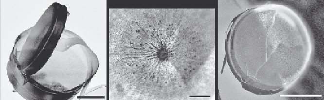
FIGURE 25.13
(a).SEM.images.of.a.complete.diatom.showing.the.two.valves.and.the.internal.structures..(b).Optical.microscope.
images. of. the. inside.of. a. valve. face. of. the. diatom,.
C. wailesii
.. The. image. shows. the. periodic. structure. that. is.
responsible.for.the.diffraction.colors.that.are.also.visible..(c).A.composite.image.of.optical.and.electron.micro-
scope.images.of.the.same.valve..The.same.features.are.clearly.visible.on.both.SEM.and.optical.images..Scale.
bars:.100.μm.(a),.20.μm.(b),.150.μm.(c).