Biomedical Engineering Reference
In-Depth Information
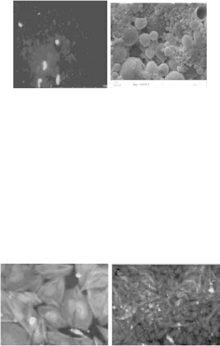
A
B
FIGURE 11.9
Fluorescence. and. SEM. micrographs. of. calcein-loaded. PHBV. nano-microcapsules.. (A). Scale. bar. is. 100. µm..
(B) 30,000×.
After.the.nano-microcapsules.of.PHBV.and.PLGA.were.stained.with.Nile.red.0.1%.(v/v),.
which. has. a. red. emission. under. luorescence. microscopy,. Saos-2. cells. were. incubated.
with.the.nano-microparticles.for.4.h..Before.examination,.the.cells.were.stained.with.FITC.
(luorescein.isothiocyanate)-labeled.phalloidin.and.4',6-diamidino-2-phenylindole.(DAPI),.
respectively,.dyes.that.stain.the.cell.nuclei.blue.and.the.cell.cytoskeleton.green..The.cells.
were.then.examined.under.luorescence.microscope.(Figure 11.10)..It.has.to.be.remembered.
that.the.sizes.of.most.capsules.are.lower.than.the.resolution.of.the.luorescent.microscope.
(in. the. nano-range),. so. some. of. the. red. regions. probably. represent. clusters. of. nanocap-
sules,.while.the.others.that.are.seen.as.individual.specks.could.be.micron.sized..It.can.be.
observed.that.PHBV.and.PLGA.nano-microcapsules.are.taken.up.by.Saos-2.cells..An.inter-
esting.inding.is.that.the.capsules.are.generally.located.near.or.on.the.cell.nuclei,.implying.
that.they.could.serve.as.carriers.of.agents.for.gene.therapy.because.they.seem.to.be.able.to.
avoid.lysosomes.and.accumulate.in.the.vicinity.of.the.nuclei.
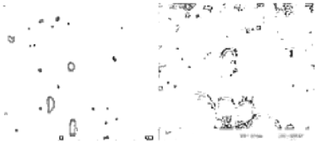
A
B
FIGURE 11.10
(See.color.insert.).Fluorescence.micrographs.of.nano-microcapsules.taken.up.by.Saos-2.cells..(A).PHBV,.40×..(B).
PLGA,.20×..Cell.nuclei.were.stained.with.DAPI.(blue),.cell.cytoskeletons.were.stained.with.FITC-labeled.phal-
loidin.(green),.and.nano-microcapsules.were.stained.with.Nile.red.(red).