Biomedical Engineering Reference
In-Depth Information
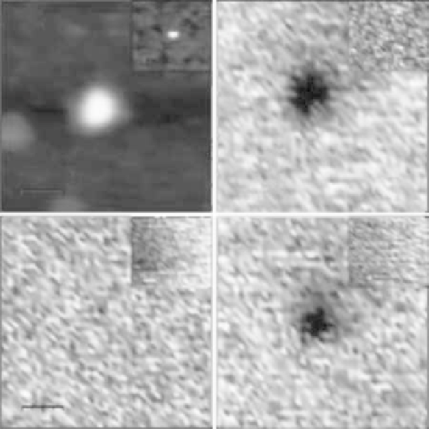
A
B
35 nm
35 nm
-5V
C
D
35 nm
35 nm
0V
+5V
FIGURE 1.3
(A).Topographic.AFM.image.of.a.single.6His-SP1-GNP.hybrid,.and.6His-SP1.with.no.GNP.attached.to.it.(inset)..
(B-D).EFM.measurements.of.a.6His-SP1-GNP.hybrid:.the.phase.shift.images.were.measured.50.nm.above.the.
set.point.height.at.negative.(-5.V).(B),.zero.(C),.and.positive.(+5.V).(D).tip.bias.voltages..The.insets.show.similar.
measurements. done. on. 6His-SP1. with. no. GNP. attached. to. it.. No. electrostatic. interaction. signal. is. observed..
(From.Medalsy.et.al..2008.)
the. 6His-SP1-GNP. hybrid. location,. for. both. positive. and. negative. bias. voltages,. is. a.
clear. evidence. for. the. existence. of. a. polarizable. object,. metallic. for. this. signal. strength,.
within.the.6His-SP1.protein..The.lack.of.electrostatic.signal.from.the.6His-SP1.(insets.in.
Figure 1.3B-D).ensures.that.the.origin.of.the.signal.is.the.GNP.bound.to.the.6His-SP1.and.
not. the. protein. itself.. The. combined. transmission. electron. microscopy. (TEM). and. EFM.
information.conirms.the.attachment.of.the.GNP.to.the.6His-SP1.protein.and.the.forma-
tion.of.the.hybrid.structure.
1.3 SP1-GNP-DNATri-BlockConjugates
One-dimensional. wires. made. of. SP1-GNP. hybrids. anchored. at. various. positions. along.
a. dsDNA. were. formed. using. a. combination. of. thiolated. ssDNA. that. partially. overlaps.
with.other.thiolated.ssDNA.and.6His-SP1-GNP.hybrids..In.these.wires.the.thiol.groups.
are.used.as.anchoring.groups.to.the.GNPs.in.the.hybrids..The.partial.overlap.between.the.