Biomedical Engineering Reference
In-Depth Information
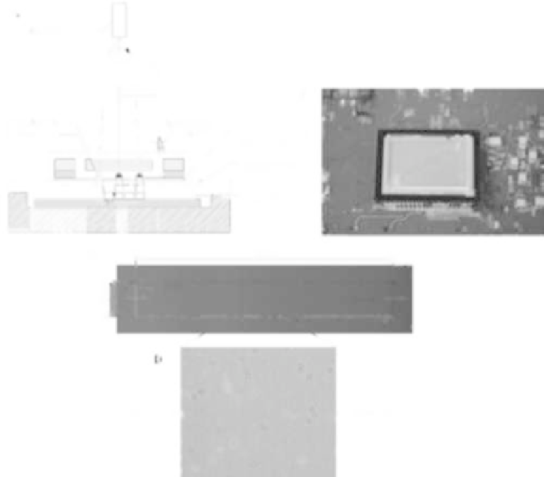
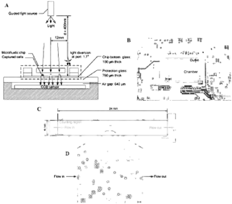
FIGURE 6.2
Schematic. illustration. of. lensless. imaging. technology.. (A). Design. of. lensless. imaging. to. detect. the. captured.
cells.in.a.microchamber..When.a.light.source.sheds.light.on.CD4.cells,.which.are.captured.in.a.microchamber,.
they.form.shadows.on.a.CCD.sensor..(B).Prototype.of.the.microchip.and.the.lensless.CCD.imaging.platform..
The.ield.of.view.of.the.CCD.sensor.is.35
× 25 mm..(C).Design.of.the.microchamber..The.microchamber.has.a.
dimension.of.4.×.24.mm..(D).The.shadow.image.of.captured.CD4.cells.in.the.microchamber.is.shown,.with.a.
scale.bar.of.100 μm..(Adapted.from.Moon,.S.,
et.al.,.
Biosens. Bioelectron.
,.24(11),.3208-3214,.2009.)
6.3.5 Single-Platform Image Cytometer (SP ICM)
Different. from. other. simpliied. low. cytometers,. a. single-platform. image. cytometer. has.
been.developed.
50-52
.This.image.cytometer.consists.of.two.major.components:.a.cell.separa-
tion.chamber.and.an.image.detection.system..To.selectively.separate.and.count.CD4
+
.cells.
from.blood,.immunomagnetic.and.luorescent.labeling.are.both.employed..T.lymphocytes.
from.whole.blood.are.irst.separated.with.immunomagnetic.nanoparticles.functionalized.
with.CD3
+
.MAb,.upon.a.homogeneous.magnetic.force..After.selective.separation.with.a.
magnet,.T.lymphocytes.are.then.labeled.with.anti-CD4
+
.and.anti-CD8
+
.MAbs,.which.are.
conjugated. to. PE. and. PerCP,. respectively.. For. luorescence. imaging,. two. light-emitting.
diodes. (LEDs). are. placed. symmetrically. with. ilters. of. 595AF60. for. PE. luorescence. and.
695AF55.for.PerCP..For.rapid.imaging,.processed.cells.are.mounted.in.an.analysis.chamber.
and.pictures.are.recorded.by.a.CCD.camera..Recent.studies.have.shown.that.this.image.