Biomedical Engineering Reference
In-Depth Information
however,.PMMA.was.used.instead.of.PDMS.due.to.its.transparence..The.selectively.cap-
tured.CD4.cells.are.counted.by.their.shadow.images.that.fall.on.a.lensless.CCD.surface.
in.a.few.seconds..Basically,.this.type.of.microchip.is.composed.of.three.layers,.including.
a.glass.coverslip,.double-sided.adhesive,.and.PMMA.base.with.an.inlet.and.an.outlet.
17
.A.
microchannel.is.formed.between.the.glass.coverslip.and.the.PMMA.base,.with.a.dimen-
sion.of.25.×.4.×.50.µm..It.is.noted.that.microchip.fabrication.can.be.easily.scaled.up.via.a.
laser.cutter..In.addition,.the.number.of.microchannels.can.be.increased.on.each.microlu-
idic.device.to.increase.the.throughput.and.the.multiplexing.capability.
6.3.2 Microscopy-Based Detection and Counting
Similar. to. low. cytometry,. microluidic-based. CD4
+
. cell. count. initially. used. luoro-
chrome.or.quantum.dot-labeled.anti-CD3
+
.and.anti-CD4
+
.MAbs,.achieving.luorescence.
detection.
23
. Captured. cells. are. viewed. through. different. luorescence. ilters. under. a.
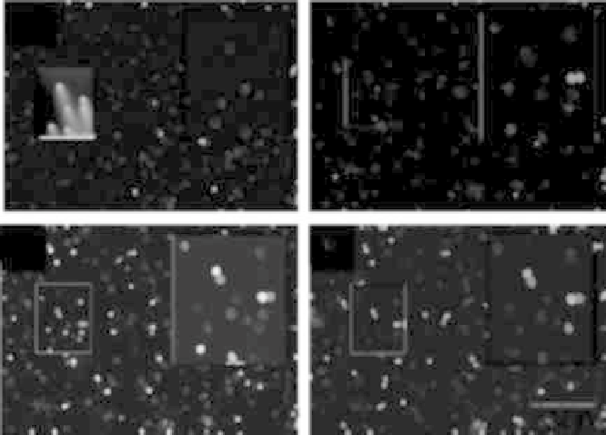
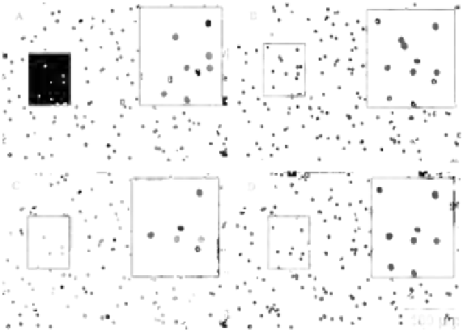
miscroscope.. As. shown. in. Figure 6.1,. CD3
+
. cells. are. shown. in. red. (panel. A). and. CD4
+
.
cells. (including. monocytes). are. shown. in. green. (panel. B).. In. a. processed. image. (panel.
C),. images. viewed. with. different. luorescence. ilters. are. digitally. overlapped,. with.
CD3
+
CD4
+
.T.lymphocytes.appearing.in.yellow..For.quantiication,.CD4
+
.T.lymphocytes.
are.counted.from.processed.images,.which.represent.CD4
+
.T.lymphocytes.in.0.18.µl.of.
FIGURE 6.1
(See.color.insert).Fluorescence.imaging.(10×).of.CD4.cells.captured.in.a.nanobiochip..(A).Cells.are.labeled.with.
anti-CD4.antibody.conjugated.with.Qdot.565.(in.green)..(B).Cells.are.labeled.with.anti-CD3
+
.antibody.conjugated.
with.Qdot.655.(in.red)..(C).CD3
+
CD4
+
.cells.are.shown.in.yellow.in.the.emerged.image..(D).Absolute.CD4.cell.count.
(in.yellow).is.obtained.in.the.processed.picture,.in.which.cells.in.red.or.green.are.deleted..A.long-pass.emission.
ilter.allows.for.CD4
+
.T.lymphocyte.counting.instead.of.image.processing..Boxes.on.the.right.in.each.panel.show.
magniied.images.from.the.left.(2×).
(Adapted.from.Jokerst,.J.V.,.et.al.,.
Lab Chip
,
8(12),.2079-2090,.2008.)