Biomedical Engineering Reference
In-Depth Information
filtered molecules. Changes within the cross-sectional area of the afferent arteriole and the
efferent arteriole can drastically affect the pressure within the capillary beds, acting as a
regulator for the filtration and absorption rates.
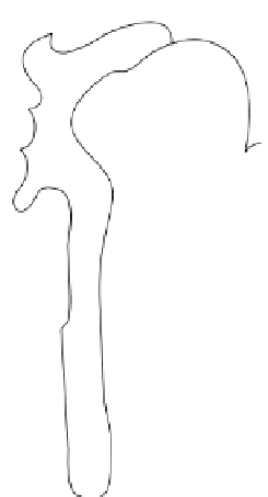
The nephrons are located within the cortex and medulla of the kidney (
Figure 12.2
).
There are nearly 2 million nephrons within the adult body, and each one has the ability to
form urine. Nephrons do not regenerate after birth; therefore, with injury, disease, and
aging there is a loss of the quantity of nephrons within the body (to about 50% of the total
quantity at the age of 80). The kidneys and the remaining functioning nephrons can adapt
to this decrease in functional unit quantity and still produce enough urine properly to
remove wastes/toxins. Each nephron is composed of two major parts. The first is the glo-
merulus, which consists of the glomerular capillaries and Bowman's capsule. The glomer-
ulus functions to filter fluid and compounds from blood. The second major component of
the nephrons is a tortuous tube which modifies the filtered fluid into urine through differ-
ential reabsorption and secretion of metabolically important molecules.
The glomerular capillaries within the glomerulus are very similar to all other capillaries
within the body, except that they are under a very high hydrostatic pressure (approxi-
mately 60 mmHg). Also, unlike other capillary beds, the glomerular capillaries are sur-
rounded by epithelial cells (termed podocytes). In one sense, these capillaries are not only
composed of a single endothelial cell wall, but they also have a second layer composed of
Proximal convoluted tubule
Bowman's capsule
Distal convoluted tubule
Glomerulus
Efferent arteriole
Cortical collecting duct
Macula densa
Afferent arteriole
Kidney's cortex
Proximal straight tubule
Kidney's Medulla
Ascending thick limb of
the loop of Henle
Descending thin limb of
the loop of Henle
Medullary collecting duct
Ascending thin limb of
the loop of Henle
FIGURE 12.2
Schematic of the functional unit of a kidney: the nephron. This figure illustrates the anatomical
arrangement of the nephron. Notice that the macula densa is a grouping of cells and not a particular region of the
kidney. Blood is filtered in the glomerulus and the filtrate passes through the nephron (proximal convoluted
tubule, Loop of Henle, distal convoluted tubule, and collecting duct) to form urine.
Adapted from Martini and Nath
(2009).


Search WWH ::

Custom Search