Biomedical Engineering Reference
In-Depth Information
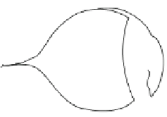
FIGURE 10.1
Anatomical structures
of the eye, showing the major optical and
sensory components. The anterior cavity
and the posterior cavity experience fluid
movement. The anterior cavity is contin-
uously forming and circulating aqueous
humor, while the posterior cavity experi-
ences a slow flow of aqueous humor.
Posterior cavity
Iris
Lens
Cornea
Pupil
Optic nerve
Anterior cavity
Retina
Sclera
attachment, and to provide protection for the interior structures of the eye. The sclera cov-
ers most of the eye structure and is composed of connective tissue (mostly collagen and
elastin). The sclera is the white portion of the eye, and it gets this color from the high con-
centration of collagen fibers present within this region. Small blood vessels and nerves are
located within the sclera. The cornea is continuous with the sclera and is located on the
anterior side of the eye, protecting it from mechanical injury and/or contamination. The
cornea also acts to protect the iris and lens of the eye. Interestingly, the cornea contains no
blood vessels and must obtain enough oxygen and nutrients through diffusion.
The second layer of the eye (middle layer) is termed the vascular tunic (or uvea), and it
contains a large quantity of blood vessels and lymphatic vessels. The primary functions of
the vascular tunic are to provide a pathway for blood vessels and lymphatic vessels, to
secrete and absorb aqueous humor (more on this in
Sections 10.2
and
10.4
), to control the
amount of light that enters the eye, and to control the shape of the lens. The blood and
lymphatic vessels are housed with the intrinsic muscle of the eye (these are smooth muscle
cells). The intrinsic muscles can partially regulate the shape of the eye through relaxation
and contraction. Also included within this layer are the iris, the ciliary bodies, and the cho-
roid. The iris is primarily composed of smooth muscle cells, which contain blood vessels
and pigment cells. When these smooth muscles cells contract, the shape and the size of the
pupil change, which effectively regulates the amount of light that enters the eye. There are
two types of smooth muscles cells, the constrictor muscles and the dilatory muscles; as the
names imply, one constricts the pupil and the other dilates the pupil. The ciliary bodies
are a specialized region of the vascular tunic that is composed of a large number of ciliary
muscles. The ligaments that are associated with these muscles are attached to the lens,
holding it in place to collect and focus all of the light that enters through the pupil. The
choroid is the name given to the purely vascular regions of the vascular tunic.
The third layer of the eye (innermost layer) is termed the neural tunic or the retina. The
inner layer of the neural tunic is termed the neural part and contains all of the photorecep-
tors of the eye. The outer layer of the neural tunic is termed the pigmented part and acts
to absorb and not reflect light that has passed through the neural part, thus preventing the
light to return into the eye structure. There are two types of photoreceptors located within
the neural part of the neural tunic. The first type of receptor is termed the rods, which are
sensitive to the intensity of light and not the wavelength (color) of the light. The rods
allow us to see structures under low light conditions. The second type of photoreceptor is


Search WWH ::

Custom Search