Biomedical Engineering Reference
In-Depth Information
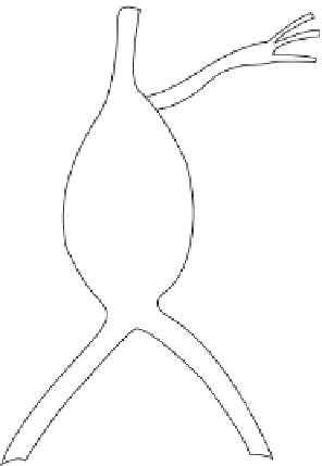
FIGURE 5.23
Schematic of a saccular aortic aneurysm, which is a
bulging of the blood vessel wall. Normally, the wall bulges because
there is a deterioration of the muscular mass in the wall and due to
the constant pressure loading the wall begins to bulge. In severe
cases, the wall can deteriorate to such an extent that the vessel breaks
and blood begins to pool in the extravascular space.
for rupture to occur increases. A blood vessel that ruptures, will cause a severe hemor-
rhage, which may eventually lead to death.
Depending on the blood vessel that has experienced an aneurysm, the effect varies.
Unfortunately, the arteries within the brain and the abdominal aorta are very susceptible
to aneurysm due to their constant high pressure loading (and unloading). A ruptured
aneurysm in the brain can cause stroke or increased pressure on the brain, while those in
the aorta will cause excessive bleeding into the chest cavity. Blood in the chest increases
the pressure on all of the organs present within the chest cavity and prevents them from
functioning properly. In either case, because the blood vessel is either supplying the brain
or some of the chest cavity organs with nutrients (depending on the location of the aneu-
rysm, although approximately 95% of abdominal aneurysms occur distal to the renal arter-
ies), the effect will be devastating.
A true aneurysm is one in which the intima of the blood vessel has bulged outside past
the media and the adventitia. A false aneurysm is caused by blood leaking and clotting in
a small space next to the blood vessel. The morphology of aneurysms varies, and the most
common have been termed saccular (resembling a sphere in three-dimensional space) or
fusiform (resembling a cylinder in three-dimensional space). Typical risk factors for aneu-
rysm include high blood pressure, diabetes, tobacco smoke, and alcohol.
END OF CHAPTER SUMMARY
5.1.
Arteries are blood vessels that transport blood away from the heart. The arterial wall is
composed of three distinct layers: the tunica intima, the tunica media, and the tunica
adventitia. The tunica intima is the inner layer of the vessel wall and is composed of endo-
thelial cells. The tunica media is composed of smooth muscle cells and is typically the
thickest layer within an artery. The tunica adventitia is composed mostly of connective


Search WWH ::

Custom Search