Biomedical Engineering Reference
In-Depth Information
2mm
2
A
5
2
51 cm
2mm
50
:
5
:
where L is the arc length of the exterior portion of the leaflet. From previous discussion, the pres-
sure in the atrium will be equal on all surfaces, including the leaflet. Therefore, the tension that
the leaflet must have to maintain its shape is
2mm
2
T
e
5
3 mmHg
50
20 mN
:
5
The tension on the inner leaflet surface is
T
i
r
i
1
T
e
r
e
p
i
2
p
e
5
T
e
r
e
20 mN
T
i
5
r
i
p
i
2
p
e
2
2
5
ð
1cm
Þ
1
5 mmHg
6mN
:
2
5
2
ð
Þ
1
:
2cm
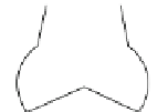
The aortic valve consists of three thin leaflets; each in a crescent shape. Immediately dis-
tal to the three leaflets there is an enlargement of the aorta (which is normally around
27 mm in diameter) termed the sinuses of Valsalva. The aortic sinus plays an important
role in valve closure, which will be discussed later. The aortic valve remains closed until
the pressure within the left ventricle exceeds the pressure in the aorta (under normal con-
ditions this is approximately 80 mmHg). Upon valve opening, the blood flow is split into
two separate streams (
Figure 4.13
). The first stream comprises the majority of the blood
and enters the ascending aorta to enter the systemic circulation. The second portion of the
blood flow is directed into the valve sinus. This blood flows in a slow vortex behind the
valve leaflets and eventually rejoins the blood in the ascending aorta, once the valve
FIGURE 4.13
Schematic of blood
flow through the aortic valve during
the cardiac cycle (the times overlap
and continue from
Figure 4.12
).
Again, the times are relative, but give
a general idea of blood flow through
the aortic valve.
600 ms
Isovolumic contraction
t
=
650 ms
Aortic valve opens
t
=
700 ms
Peak systole
t
=
t
=
750 ms
t
=
800 ms
t
=
850 ms through diastole
Aortic valve closed


Search WWH ::

Custom Search