Biomedical Engineering Reference
In-Depth Information
The goal of this discussion is to illustrate how solid mechanics and fluid mechanics can
be coupled to determine the pressure forces that are acting on blood within the heart and
that can even drive fluid flow through the heart and the cardiac vasculature. For a course
at this level, to calculate a solution, many assumptions need to be made concerning the
geometry, the mechanical properties, and the fluid properties. With these assumptions, a
meaningful solution can still be reached (see Bernoulli Equation, Section 3.8), but it is
more accurate to minimize the assumptions made. As stated before, solutions that use this
complex approach will be discussed in Chapter 13.
4.5 HEART VALVE FUNCTION
As discussed in previous sections, the primary function of the heart valves is to prevent
the backflow of blood during muscle mass contraction. The mechanism by which the
valves accomplish this is fairly interesting, and this section will briefly discuss valve func-
tion. We will discuss the mitral valve and the aortic valve, because this is where most of
the research has been conducted. The function of the tricuspid valve mimics the mitral
valve and the function of the pulmonary valve mimics the aortic valve (again, recall that
the pressures will be lower on the right side of the heart). The anatomy of heart valves is
shown in
Figure 4.11
and is discussed in the following section.
Recall that the mitral valve regulates the blood flow between the left atrium and the left
ventricle. The mitral valve consists of two thin membranes, which are connected by ten-
dons (the chordate tendineae) to the papillary muscles. The papillary muscles in turn are
connected to the left ventricular muscle mass. The mitral valve remains open, allowing
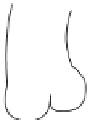
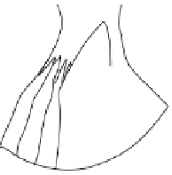
FIGURE 4.11
The mitral valve (A) and the aortic
valve (B) determine the blood flow characteristics and
blood flow direction through the left side of the heart.
The chordate tendineae and the papillary muscles do not
open or close the mitral valve, but instead prevent the
leaflets from bulging into the atria during ventricular con-
traction. The coronary arteries come off the aortic valve
sinuses and feed the cardiac tissue with blood.
A
Chordate tendineae
Papillary muscles
Side view
Top view
Aorta
B
Coronary arteries


Search WWH ::

Custom Search