Biomedical Engineering Reference
In-Depth Information
system that drives the blood flow throughout the body or lungs. In the next few sections,
we discuss the physiological controls on heart motion and how blood flows through the
heart. Blood flow through the vascular system will be discussed in subsequent chapters.
To understand the physiology of the heart and how the heart functions, we first need to
understand what cardiac muscle is and how it functions. Cardiac muscle cells (or cardiac
myocytes) are interconnected in a very special way, allowing the cells to function as one.
This is dissimilar to skeletal muscle, which readers may be more familiar with from previ-
ous physiology courses. Individual cardiac muscle fibers continually divide and merge,
forming a highly interconnected mesh-like structure (
Figure 4.2
). The cell membranes of
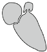
FIGURE 4.1
Anatomy of the heart
depicting the direction of blood flow
through the four heart chambers. The
right side of the heart collects deoxy-
genated blood from the systemic circu-
lation and delivers it to the pulmonary
circulation. The left side of the heart
collects oxygenated blood from the pul-
monary circulation and delivers it to
the systemic circulation.
Aorta
Pulmonary
artery
Lungs
Superior
vena cava
Pulmonary
vein
Left atrium
Right atrium
Left ventricle
Right ventricle
Inferior vena
cava
FIGURE 4.2
Schematic of car-
diac muscle cells depicting the
highly interconnected fibers that
form a syncytium, and the interca-
lated discs that divide individual
muscle cells. There are many gap
junctions present within the inter-
calated discs to facilitate the
movement of ions between cells,
thus allowing the action potential
to continuously flow from one cell
to the next. Adapted from Guyton
and Hall (2000).


Search WWH ::

Custom Search