Biology Reference
In-Depth Information
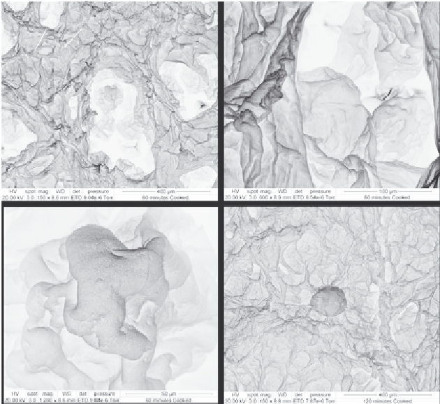
A
B
Wrinkled cell wall components
C
D
Figure 4.9 Scanning electron micrographs taken during in vitro small intestinal diges-
tion of starch in Agria cooked potatoes. (A-C) Samples taken after 60 min (at different
magnifications) and (D) 120 min. As artifacts produced by freeze drying are present,
these micrographs do not necessarily represent the structure prior to freeze drying.
Reproduced from
Bordoloi, Singh, et al. (2012)
with permission from Elsevier.
cooked potato. The regular microcellular structure of cooked potato tuber
appeared to have been maintained to a good extent during and after cooking.
A good level of cell integrity was observed in all the samples. The tuber cells
appeared to have shrunk slightly and showed indentation and wrinkles on
their surface. The cell wall of the undigested sample showed less wrinkles
compared to the samples subjected to small intestinal digestion. Hydrolysis
of starch during
in vitro
digestion and removal of water during freeze drying
might have left an empty space in which the cell wall and some undigested
components could have folded in during freeze drying, causing more wrinkles
and indentation. This phenomenon was especially distinctive in cells that
underwent the complete 120-min simulated intestinal digestive process
(
Fig. 4.9
D). However, the possibility of some artifacts produced due to freeze