Biology Reference
In-Depth Information
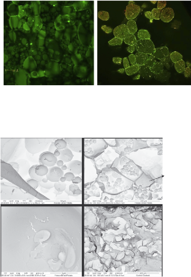
Figure 4.2 Confocal laser scanning micrographs of raw (left) and cooked (right) tuber
parenchyma from Agria potato cultivar. Reproduced from
Bordoloi, Kaur, et al. (2012)
with
permission from Elsevier.
Immature starch
granules
A
B
Cellular
cytoplasm
Unopened cell
C
D
Cytoplasmic
starchy matrix
Figure 4.3 SEMmicrographs of raw (A-C) and cooked (D) tuber parenchyma from Agria
potato cultivar. In raw potato parenchyma micrographs, starch granules are seen
embedded in the cellular cytoplasm. Indentations and pores on the surface of starch
granules are clearly seen along with other cellular remains and tiny granular structures
resembling “immature starch granules” as reported by
Singh et al. (2005)
. Please note
that the cracks on the starch granules might have formed during microscopy.
Reproduced from
Bordoloi, Kaur, et al. (2012)
with permission from Elsevier.