Biomedical Engineering Reference
In-Depth Information
3500
3000
2500
Chloride
Bromide
Iodide
2000
1500
1000
500
0
0.0
0.5
1.0
1.5 2.0 2.5
Salt concentration (m)
3.0
3.5
4.0
FIGURE 19.5
Attenuation increases associated with increasing iodine concentration. In general, a change of 50-100 HU (Hounsfield Units) is
more than adequate to perceive a difference in contrast. It is apparent that even low concentrations of iodide are readily detected and higher con-
centrations effectively saturate the system.
that contrast, reagents, and salt products would migrate through
tissues at different rates.
To address this we considered that it should be possible to use
an acid with the anion positioned lower on the periodic table
with a higher atomic number and atomic mass. Two obvious
choices were HBr and HI. Both of these are strong acids, and
when tested in the phantom, performed equally as well as their
less dense counterpart, HCl, at the maximum concentrations
available. In particular, HI is attractive because it has a dramatic
effect on attenuation at CT even at relatively low concentrations,
as shown in Figure 19.5, where the relative attenuation increases
with the increase in iodine content.
Thus, it is possible to use a portion of HI mixed with HCl to
obtain a suitably dense mixture for thermochemical ablation.
Unlike adding contrast, which has a very different structure and
physical properties than the acid, it seems safe to assume that
where the density from iodide is present, there too is acid.
The next logical step was
in vitro
testing in tissues. In order
to do this, though, it was necessary to invent a device that would
channel two reagents into the tissues but only allow reaction at
or very near the tip. The resources for a custom device were not
available, and furthermore we considered it desirable for others to
be able to repeat the results elsewhere without a large investment.
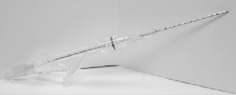
A survey of existing components readily available in the interven-
tional radiology suite led to a combination shown in Figure 19.6.
The components are a coaxial biopsy cannula/trocar system,
a rotary hemostatic valve attached to the hub, and a smaller
coaxial needle. The needle must be of sufficient length to pass
through the valve such that the tip extends nearly to the tip of
the cannula. There are then two injection ports, one through the
inner needle, and one via the side arm of the hemostatic valve.
It is relatively straightforward to connect these via extension
tubing to syringes loaded with reagents, and a syringe pump can
be incorporated to ensure consistent injection rates.
Use of such a device then allowed us to test thermochemical
ablation in tissues such as
ex vivo
liver. We used a combination
of thermocouple data and infrared imaging for the initial set of
experiments.
35
Although infrared imaging is not capable of pen-
etrating deeply into tissues and is not a volumetric modality, it
did provide the first level of data upon which to build. Sectioned
tissues are shown in Figure 19.7 to demonstrate the shape of
the heated area at least on the exposed surfaces, in which the
specimens were bivalved as soon as possible after the comple-
tion of injections to visualize the heated area. Here again, as in
the
in vitro
phantom experiments, the chemistry was consistent.
FIGURE 19.6
Components of a prototype thermochemical ablation
device. A rotary hemostatic valve is connected to a biopsy outer can-
nula, and through this assembly a long needle is inserted coaxially such
that the tips are at nearly the same distance. When the two injection
ports are connected to appropriate solutions via tubing, the reagents
will only mix and react near the tip, exiting as a hot, hyperosmolar
solution.