Biomedical Engineering Reference
In-Depth Information
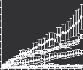
enhancement of extravasation in tumors between 39 and 40°C
(Kong et al. 2001). The rate of extravasation doubled for each 1°C
temperature rise, up to a maximum temperature of 42°C (Figure
16.2b). Above this temperature, hemorrhage obscured the ability
to observe further thermal effects on extravasation in the mod-
els that he used.
Kong also found evidence for enlarged pores up to 4 hr post
heating, but by 6 hr, they had closed to pre-heating levels. When
he attempted to reopen them by a second heat, delivered 8 hr
after the first heating, they did not reopen. This provided evi-
dence that the closing of the pores was likely associated with the
heat shock response and in effect was a manifestation of thermo-
tolerance (Kong et al. 2001).
To examine whether hyperthermia would augment liposomal
accumulation in a spontaneous tumor, Matteucci et al. radiola-
belled a long-circulating liposomal drug formulation and exam-
ined tumor uptake of these liposomes with a gamma camera in
pet cats with spontaneous soft tissue sarcomas. Baseline scans
were performed without heating and then followed with heat-
ing several days later in the same animals. Similar to what was
seen in rodent models, hyperthermia enhanced the uptake of
liposomes by factors between 2 and 13 fold (Matteucci et al.
2000). Kleiter later determined that the relative enhancement of
liposomal uptake after hyperthermia treatment, as observed by
gamma camera in rats with transplanted mammary carcinomas,
was linearly proportional to doxorubicin uptake for sterically
stabilized Doxil
•
liposomes (Figure 16.3) (Kleiter et al. 2006).
Goldberg examined the effects of thermal ablation on accu-
mulation of the pegylated liposome Doxil
•
, finding that RF abla-
tion administered prior to Doxil
•
administration led to greater
than a five-fold increase in drug accumulation, with the greatest
concentrations being seen at the periphery of the ablation zone.
It is likely that the enhanced liposome accumulation observed
with this combination therapy is the result of the combination
34ºC
1 minute
30 minutes
60 minutes
42ºC
(a)
2.0
1.8
1.6
1.4
1.2
1.0
0.8
0.6
0.4
0.2
0.0
2.0
1.8
1.6
1.4
1.2
1.0
0.8
0.6
0.4
0.2
0.0
39
40
Te mperature (°C)
41
42
0510 15 20 25 30
Time (min)
35 40 45 50 55 60
(b)
FIGURE 16.2
(a) Depiction of liposomal extravasation from tumor microvessels at 34 vs. 42°C, up to 60 min of heating. Data acquired using
intravital microscopy of SKOV3 tumor microvessels in skin-fold window chambers. The liposomes were fluorescently labeled, which permitted
visualization of the tumor microvessels as well as the extravasation of the liposomes, which can be visualized as bright perivascular spots of accu-
mulation. (From Kong, G., Braun, R. D., Dewhirst, M. W.,
Cancer Res
, 60, 2000. With permission.) (b) (left panel). Rate of liposomal extravasation
from SKOV3 tumor microvasculature, as a function of temperature. • = 42°C, ∗ = 41°C, π = 40°C, lowest sets of curves represent 34 and 39°C,
respectively. (right panel). Relative amount of interstitial liposomes as a function of temperature. The slope of this curve predicts that the accumu-
lation of liposomes doubles for each 1°C temperature rise. (From Kong, G. et al.,
Cancer Res
, 60, 2000. With permission.)