Biomedical Engineering Reference
In-Depth Information
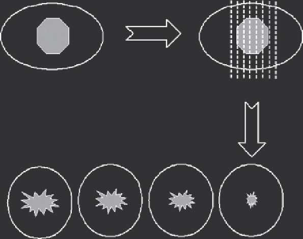
US Imaging divides the tumor
into slices (from 1 to
N
)
US Imaging
Tumor
US Slice 1 2 3...
N
US Imaging
Slice 4
US Imaging
Slice 3
US Imaging
Slice 2
US Imaging
Slice 1
FIGURE 15.1
Schematic diagram showing that the entire tumor can be segmented into slices with ultrasound imaging.
15.5 HIFU 3-D Conformal therapy
A single exposure can be made to induce a cigar-shaped
lesion when the location of the focal volume is immoveable.
The exposure time for each pulse ranges from one second to
several seconds. The single exposure can be repeated at pre-
determined intervals in the same position. Multiple single
exposures can produce a line-shaped lesion by placing single-
exposure lesions side by side with a present overlap and with a
predetermined time interval between exposures. The multiple
single exposures can be repeated in the perpendicular direc-
tion to form a slice-shaped lesion. A line-shaped lesion can also
be achieved by a linear track exposure in which the activated
transducer is moved at a constant speed over a line. This may
be made by traversing one or more times in one direction only,
or by scanning in both directions (“there and back”) without
pausing at the furthest extremity. Several of these tracks may
be superimposed in one exposure period at chosen (preset) time
intervals. Superimposition of tracks leads to an increase in the
extent of ablation in the direction perpendicular to the direction
of motion due to thermal conduction. As a result, a slice-shaped
lesion can be induced in this way. From one slice-shaped lesion
to the next, confluent volumes of ablation can be achieved.
The selection of the previous exposure regimes during HIFU
procedure is very complicated in clinical practice. It depends on
the component of overlying tissue structure, acoustic window,
the depth of tumor from the skin, vital structures surrounding
tumor, tumor vascularity, and size. For instance, single expo-
sure and a linear scan exposure can be chosen for the treatment
of a superficial, poorly vascularized tumor. However, multiple
single exposures are usually used in the treatment of deep vas-
cularized tumors. In clinics they can be separately used for an
The volume of tissue ablation induced with one HIFU expo-
sure can be regarded as a lesion. The focal volume of a HIFU
transducer is usually ellipsoid or cigar shaped, with dimen-
sions of 10-20 mm along the beam axis and 1-2 mm in the
transverse direction. Therefore, the lesion induced with a
single HIFU exposure is usually very small. But, while lesions
are placed side by side, confluent volumes of ablation can be
achieved.
To ablate clinically relevant volumes of tissue for the treat-
ment of carcinomas, many of these small lesions should be
positioned side by side systematically to “paint out” or cover a
targeted volume, without any remaining live tissue between each
lesion. If a suitable therapy plan is correctly performed, HIFU
is able to ablate various shapes and sizes of solid tumors in a
conformal fashion.
As a result, conformal HIFU therapy can be clinically defined
as a precise procedure to ablate an entire tumor by moving high-
energy concentrated focus side by side in a 3D fashion. At the
beginning of a HIFU procedure, the targeted tumor is identified
and divided into parallel slices of 5-mm separation by moving the
diagnostic probe. Using HIFU exposure regimes, the tumor on
each slice is completely ablated, and this process is repeated slice
by slice to achieve complete coagulation of the targeted tumor, as
shown as Figure 15.2. There are four exposure regimes that are
usually used in HIFU procedure. They include single exposure for
a cigar-shaped lesion, either multiple single exposures or linear
scan exposures for a line-shaped lesion, and a convergent scan for
the lesion of deep tumor.