Biomedical Engineering Reference
In-Depth Information
(b)
3D axial motion: tc115 frame 15
(a)
Motion field
0.2
15
0.1
10
0
5
−0.1
0
−0.2
30
Left top
Right top
Left bottom
Right bottom
20
−0.3
15
10
10
−0.4
Lateral (mm)
5
36
38
40
42
44
46
0
0
−5
Elevation (mm)
Temperature (°C)
(c)
3D lateral motion: tc115 frame 15
(d)
3D elevation motion: tc115 frame 15
0.2
0.2
0.1
0.1
0
0
−0.1
−0.1
−0.2
−0.2
−0.3
−0.3
−0.4
−0.4
36
38
40
42
44
46
36
38
40
42
44
46
Temperature (°C)
Temperature (°C)
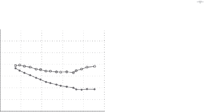
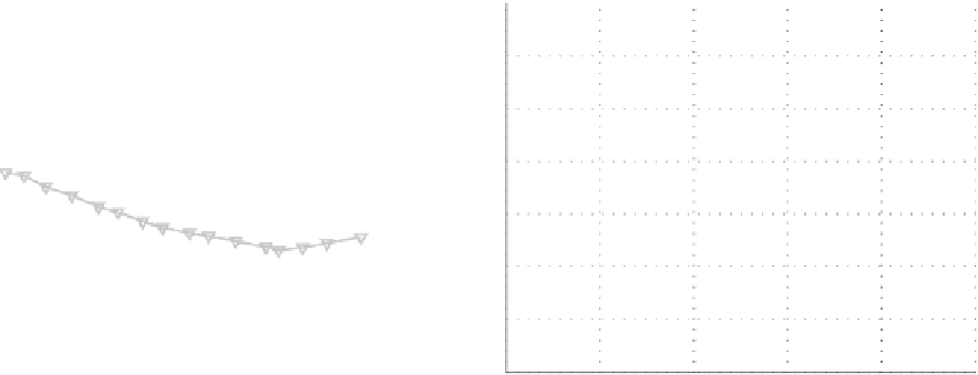
FIGURE 13.4
Accumulated nonrigid motion estimation in 3D over a 5 cm
3
volume. (a) Typical 3D motion field with 5 × magnification at the cor-
ners of the tissue volume. (b) Axial, (c) lateral, and (d) elevation displacements for one specimen of turkey breast muscle at the center image (#15)
of the 3D image set. Displacement values were found via trilinear interpolation of the estimated nonrigid 3D motion field over the image volume.
he magnitude of the motion in a 0.5°C step was <20μm (similar to Figure 13.3 in Arthur et al.
73
).
13.4 Ultrasonic Measurement
of temperature
Temperature dependence of ultrasonic tissue parameters has
been reported extensively from
in vitro
analyses of properties
that characterize tissue response to insonification.74-84
74 -84
hese
early investigations reported changes in tissue characteristics
with temperature in order to evaluate thermal errors in tissue
characterization. Some investigators did consider, however, the
possibility of using temperature dependence of tissue properties
as a means to track temperature changes.
76, 77, 85
The primary ultrasonic parameter examined for its depen-
dence on temperature in early work on measurement of tem-
perature was SOS.
77,80,85
In these initial studies investigators
tried to obtain SOS maps of the medium from which to infer
temperature distributions. This approach, however, has never
been instituted clinically.
86
Perhaps this approach has not been
implemented because in order to measure SOS, it is necessary
to measure both distance and time to image an identifiable
target from two directions, or to use a crossed-beam (multiple
beams) method.
87
Such measurement is further complicated
by the fact that ultrasonic windows do not always exist
in vivo
to allow insonification of a region of interest from two views.
Another problem is that the temperature dependence of SOS
differs depending on the tissue type (e.g., whether tissue has
high water or fat content.)
More recently, the use of ultrasonic parameters as a guide
for thermal therapy has been revisited with several differ-
ent parameters being considered. In particular, papers by Sun
and Ying;
35
Seip, Simon, Ebbini, and coworkers;
59,60,88
Maass-
Moreno, Damianou, and coworkers;
58,89,90
and our group
42,70,91
have reported the changes in received ultrasonic signals due to