Biomedical Engineering Reference
In-Depth Information
prolong survival. TACE relies on inherent differences in blood
supply to the hepatic tumor versus the normal hepatic paren-
chyma. Because hepatic tumors are supplied almost exclusively
by the hepatic artery, high intratumoral chemotherapeutic con-
centration with low systemic exposure can be achieved through
transcatheter arterial infusion into the vessels directly supply-
ing the tumor. Recent developments in TACE include the use
of drug eluting microspheres that provide simultaneous delivery
of chemotherapy and embolization with controlled drug release
over time (Liapi et al. 2010).
It should be noted that TACE is not a thermal therapy, how-
ever we have included it in this chapter as it is an important
technique in the interventional oncology armamentarium, and
the planning systems that have been developed for TACE as
described following might be applied to guidance of thermal
therapies in the future. In addition, we will describe the role of
TACE in combination therapy in Section 12.4.
While navigation techniques have not been directly applied
to TACE, there have been some reports of planning systems
that have been developed to identify the vessels feeding a
tumor or to evaluate TACE treatments (Deschamps et al. 2010).
In a study of 18 consecutive patients undergoing TACE, the
tumor feeding vessels were identified using three-dimensional
analysis on a General Electric Advantage Workstation (Figure
12.11). The images were acquired on a General Electric Innova
rotational angiography system that provides cone-beam CT
imaging using a flat panel detector. The determination of feed-
ing vessels was done by postprocessing of the images in three
steps: (1) structure extraction; (2) target definition; and (3)
feeding vessel selection. Structure extraction is accomplished
by manual placement of a seed point at the entrance of the
hepatic artery and applying thresholding and intensity-based
segmentation to “grow” the vasculature. For target definition,
the user adjusts a region of interest around the tumor. The
feeding vessels are then highlighted, and background struc-
tures such as the spine can be hidden for better visualization.
The study conclusion was that vessel segmentation and three-
dimensional analysis has a higher sensitivity in determining
sub-segmental feeding vessels than using two-dimensional
imaging alone.
In a related study, contrast-enhanced ultrasound was used
for postinterventional follow-up after TACE (Ross et al. 2010).
The ultrasound images were fused with CT or MRI of the liver
after treatment to evaluate the tumor vascularity and perfusion
of HCC tumor lesions. The study conclusion was that image
fusion provided a better visualization of the microcirculation
and the residual tumor perfusion at an earlier time point than
the usual modalities such as non-contrast enhanced CT. While
there was no real-time navigation used during the procedure,
both this study and the preceding study showed the value of
providing more imaging information during the procedure,
and one might speculate that navigation during the procedure
could help as well.
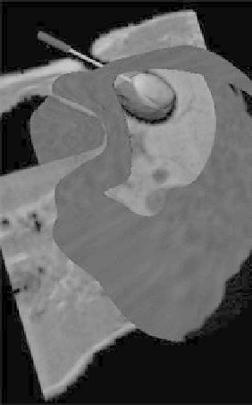
FIGURE 12.10
MR image in plane with the cryoprobe during a por-
cine study, showing the artificial tumor colored according to the tem-
perature, and the surface of the liver. (Reprinted from Samset, E., Mala,
T., Aurdal, L., and Balasingham, I.,
Comput Med Imaging Graph
29,
2005. © 2011, with permission from Elsevier.)
fibroids, and three metastatic liver tumors (Mogami et al. 2002).
The MRI system was a 0.3T AIRIS II (Hitachi, Tokyo, Japan).
The optical navigation system was based on a Polaris tracking
system and a passive reference frame that was attached to the
distal end of the cryoprobe. The size of the lesions ranged from
1.2 cm (metastatic liver tumor) to 9.0 cm (uterine fibroid). The
cryoprobes used ranged 2 to 3 mm in diameter, and the number
of probes used range from one to three. The navigation system
was considered useful in the precise placement of the probes,
although some disadvantages were noted, including the need to
maintain a line of sight from the tracking system to the reference
frame and the fact that needle bending cannot be accounted for
since the tip of the cryoprobe is not tracked.
A team from Brigham and Women's Hospital in Boston
looked at image registration of preprocedural MRI and intra-
procedural CT to visualize renal tumors during CT-guided
cyroablation procedures (Oguro et al. 2011). Both rigid and non-
rigid registration methods were used. The registration methods
were applied retrospectively to 11 CT-guided procedures, and
the team concluded that the nonrigid method was more accurate
and could be used to improve visualization.
12.3.4 transarterial Chemoembolization
Transcatheter arterial chemoembolization (TACE) is a mini-
mally invasive approach for treatment of unresectable hepato-
cellular carcinoma (HCC) that may provide symptom relief and