Biomedical Engineering Reference
In-Depth Information
Xli-e semi-
flex probe
1500X RF generator
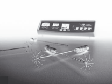
Starburst® Xli-e RFA probe
(a)
(b)
Cool-tip
TM
RF probe
Cool-tip
TM
RF generator
with chilled water pump
(c)
FIGURE 12.6
Images of three RFA devices for soft-tissue application: (a) Boston Scientific RF3000 (With permission, © 2011 Boston Scientific
Corp.); (b) AngioDynamics StarBurst
•
devices shown with multi-tined LeVeen
•
probes (With permission, © 2011 AngioDynamics
•
); (c) Valleylab/
Covidien Cool-tip
TM
device. (With permission, © 2010 Covidien.)
necrosis in a more complete, predictable, and spherical manner.
The tines can also be partially deployed to adjust the size of the
necrotic volume.
The major U.S. Food and Drug Administration (FDA)-
cleared RFA devices in use for soft-tissue ablation include the
RF3000 device (Boston Scientific, Natick, MA), the StarBurst
•
Talon device (AngioDynamics, Latham, NY, previously RITA
Medical Inc.), and the Cool-tip
TM
RF device (Valleylab, Boulder,
CO, now a subsidiary of Covidien). Multiple-tine LeVeen
•
probes are available for Boston Scientific and AngioDynamics
systems. These devices, along with their respective probes, are
shown in Figure 12.6.
applications, mirroring clinical practice. Both optical and elec-
tromagnetic tracking systems have been investigated, as well as
CT and MRI guidance. While several studies were simulations
based on clinical data sets, a few studies have incorporated
navigation systems in clinical practice. Each of the studies
listed in the table will now be described. One of the earliest
groups to investigate navigation for RFA developed a 3D simu-
lator and treatment planning system for optimal needle place-
ment (Villard et al. 2005). The system included segmentation
of the liver and the tumor, and allowed the user to virtually
place the radiofrequency probe and observe a meshed spheroid
representing the 60°C isosurface. The system also included the
ability to simulate the placement of multiple needles to treat
larger tumors and the optimal positioning of each needle to
fully treat the lesion while minimizing damage to vital struc-
tures. Simulations were completed using clinical data from 12
patients having a single, small liver tumor.
The issues with current radiofrequency ablation techniques
and the need for accurate image guidance were described by
12.3.2.2 Navigation Systems and rFa
Several researchers have investigated the use of image-guided
navigation to assist in accurate placement of the radiofre-
quency ablation probe. The major studies investigating the
use of navigation systems and RFA are shown in Table 12 .1
below. From the table, it can be seen that the focus is on liver