Biomedical Engineering Reference
In-Depth Information
Biological behavior
Histopathological exam
Lesion range
Correlation between the lesion and
Real-time adjustment
surrounding normal tissue
Optimal beam pathway
Distance from deeper layer to the skin
Real-time
monitoring
Adjuvant therapies
Selection of transducer
3D conformal scanning
Delivery of therapeutic dose
Imaging exams like CT/MRI
erapy
TPS
Blood circulation of the lesion
Effect
evaluation
and the organ
Doppler
Systemic situations
Adjustment
Physical exam
Other therapies
Review the patient's history
FIGURE 10.12
Development of the therapeutic plan system (TPS) for HIFU clinical application.
According to the surgical standards for tumor resection, it
is important to obtain safe margins. The extent of the treated
volume may vary because of different therapeutic purposes.
Additionally, the margin size is also dependent on the histologi-
cal type of tumor. Therefore, a 3D conformal HIFU treatment
plan includes not only ablation of the tumor but also surround-
ing normal tissues in order to achieve the proper margin.
10.3.5 treatment Dose Delivery
The measurement of the dose required for successful HIFU
radiation is a controversial and evolving topic. It is difficult to
quantify the biological effects of HIFU on living tissues by using
ultrasonic energy deposited in the targeted tissue and the expo-
sure time, or by theoretical calculations. Based on a large amount
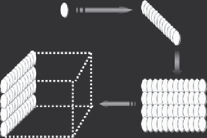
Spot
Line-shaped ablation
Spot-shaped lesion
Line-shaped lesion
Volume-shaped ablation
Slice-shaped ablation
Slice-shaped lesion
Volume-shaped lesion
(a)
(b)
FIGURE 10.13
(a) Schematic diagram shows the conformal therapeutic plan for HIFU therapy, whose 3D treatment strategy is composed of spot,
line, slice, and volume ablations; (b) after HIFU exposure, the spot-shaped, line-shaped, slice-shaped, and volume-shaped coagulation necrosis in
ex vivo
bovine liver.