Biology Reference
In-Depth Information
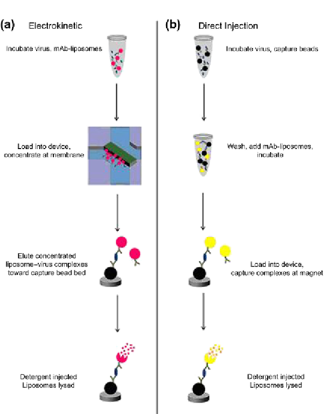
Figure 10.6
Virus detection using liposomes. Source:
Figure 1 from Ref.
82
.
Schematic
of assays with and without preconcentration. The assay employing electrokinetic pre-
concentration (a) begins with loading the device with antivirus pAb-labeled Protein A
superparamagnetic beads to create a capture bed and incubating the antivirus mAb-
labeled fluorescent liposomes with virus. The sample is then loaded into the inlet well,
concentrated at the nanoporous membrane, and eluted toward the capture bead bed.
Following washing, detergent is injected to lyse the liposomes, releasing the fluorescent
dye for quantification. The assay without preconcentration (b) begins with incubating
a virus sample with the same capture beads as before. The virus-bead complexes are
washed and incubated with electrochemical liposomes. This sample is pulled into the
microfluidic channel where the detection complexes are captured by a magnet and
washed, and the bound liposomes are lysed with detergent. This releases the electroac-
tive species, which undergoes redox cycling at a downstream IDUA. (For color version of
Search WWH ::

Custom Search