Biology Reference
In-Depth Information
they can be observed through a microscope. The company also developed
automated image recognition software for identification of the samples.
Staining can be done by flowing the dye in the channel, the circular chip
can be rotated to allow automated inspection of all channels, and the filter
membrane plug can be removed for further inspection.
Miniaturization of optical microscopes also presents a way forward for
cheaper and more portable instruments. In 2011, U.S. researchers developed
an integrated microscope that weighed just 2g and was slightly bigger than
the end of a pencil,
16
illustrated in
Fig. 5.4
. For waterborne pathogens, in 2010,
Mudanyali et al. built a portable holographic microscope and developed a rapid
image reconstruction algorithm as well as an automated counting method. An
incoherent light source was used by the lightweight microscope to illuminate
the sample of interest, while a complementary metal -oxide -semiconductor
(CMOS) chip acquired holographic images of the sample in a 60µL sample
volume. An LOD of 380cystsmL
−1
was reported for
Giardia lamblia
and the
automated counting algorithm proved capable of distinguishing between
Cryp-
tosporidium parvum
,
G. lamblia
, microbeads and dust particles. Without precon-
centration, the system was incapable of accurately detecting 189cystsmL
−1
,
although this could be improved with the use of sample-processing techniques.
18
A 2012 paper showed how a cell phone camera could be used to detect
E. coli
to an LOD of ∼5-10 cfu mL
−1
in a fluorescent assay where the excita-
tion light was provided by battery-powered inexpensive LEDs. Specificity
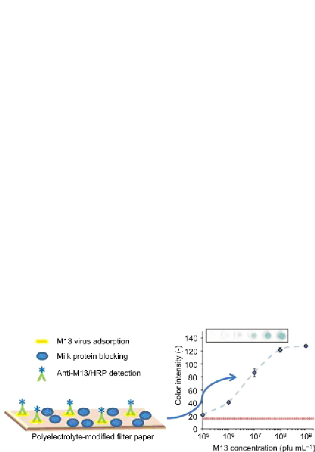
Figure 5.3
Paper-based colorimetric detection of viruses. Left: Schematic of the detec-
tion scheme. Right: Range and detection limits of the assay using a sample volume of
0.50 mL at pH 5. Values are means of six discs given with 95% confidence limits. The par-
allel lines indicate background intensity and corresponding confidence limits. The inset
shows scanned sample discs (10
5
-10
9
), including a background disc (no phage; far left).
Source:
Graphical abstract from Ref.
17
. Reproduced with permission.
(For color version of
this igure, the reader is referred to the online version of this topic.)
Search WWH ::

Custom Search