Biology Reference
In-Depth Information
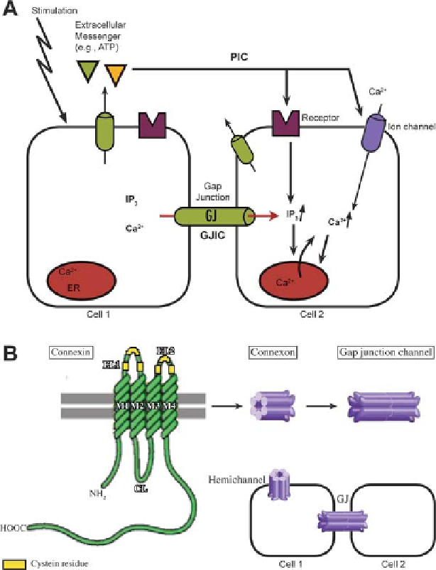
Fig. 10.1
(
a
) Mechanism of the Ca
2+
wave propagation between cells. A specific stimulus induces
aCa
2+
transient in the stimulated cell (cell 1) via Ca
2+
influx and/or Ca
2+
release from intracellular
stores (ER). Diffusion of Ca
2+
or IP
3
through the gap junctions (GJ) or secretion of an agonist into
the extracellular space that diffuses to neighboring cells can induce a Ca
2+
increase in the neighbor-
ing cells, resulting in gap junctional intercellular communication (GJIC) or paracrine intercellular
communication (PIC), respectively. (
b
) Structure of connexin, connexon hemichannels and gap
junction channels.
Top
: One connexin molecule, showing the four typical transmembrane domains
(M1-M4), together with the one cytoplasmic (CL) and the two extracellular (EL1 and EL2) loops.
The N- and C-terminal domains face the cytoplasm and are very divergent between connexins. Six
connexins form a connexon, which can be homomeric or heteromeric, depending on the connexin
isoform composition of the connexon. Connexons from adjacent cells can dock to form gap junc-
tion channels. Each cell can contribute the same or a different type of connexon, giving rise to either
homotypic or heterotypic intercellular channels. Connexons can also be present in non-junctional
membranes as connexon hemichannels
Search WWH ::

Custom Search