Biology Reference
In-Depth Information
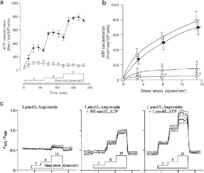
Fig. 9.7
Effect of angiostatin on shear stress-dependent ATP production and calcium influx in
HPAEC. (
a
) Extracellular ATP generation by HPAEC was measured under static conditions (
◦
)
and in response to a stepwise increase in shear stress (
). ATP concentration increased in a dose-
dependent manner with the rise in shear stress. (
b
) Extracellular ATP generation was measured
after a one-hour incubation in the presence of media alone (
•
◦
), 0.5
μ
mol/L angiostatin (
),
or 1.0
mol/L angiostatin (
). Treatment with angiostatin caused a decrease in shear stress-
dependent ATP synthesis that was reversed by flushing away the angiostatin (
μ
). The effect of
angiostatin indicates that ATP synthase is responsible for the shear stress-dependent ATP genera-
tion. (
c
) Changes inintracellular Ca
2+
levels of HPAEC in response to shear stress were measured
using the ratiometric Ca
2+
indicator indo 1-acetoxymethyl ester. Relative Ca
2+
levels are indi-
cated by the 405-to-480-nm fluorescence ratio (F
405
/F
480
). In the presence of angiostatin, a shear
stress-dependent rise in intracellular Ca
2+
does not occur. Addition of exogenous ATP restores
the dose-dependent rise in shear stress-induced Ca
2+
levels normally observed in the absence of
angiostatin. (Reprinted with modification from Yamamoto et al. [51], used with permission of the
American Physiological Society [51])
•
implicated in endothelial responses to shear stress [49, 51]. Yamamoto et al. demon-
strated that flow-induced shear stress causes an influx of calcium into human
pulmonary artery endothelial cells (HPAEC) that is largely dependent on an increase
in extracellular ATP level. Cultured HPAEC release ATP in response to shear stress
in a dose-dependent manner (Fig. 9.7a). Similar results have been observed in
freshly-isolated endothelial cells from rabbit thoracic aorta [6]. Degradation of ATP
Search WWH ::

Custom Search