Biology Reference
In-Depth Information
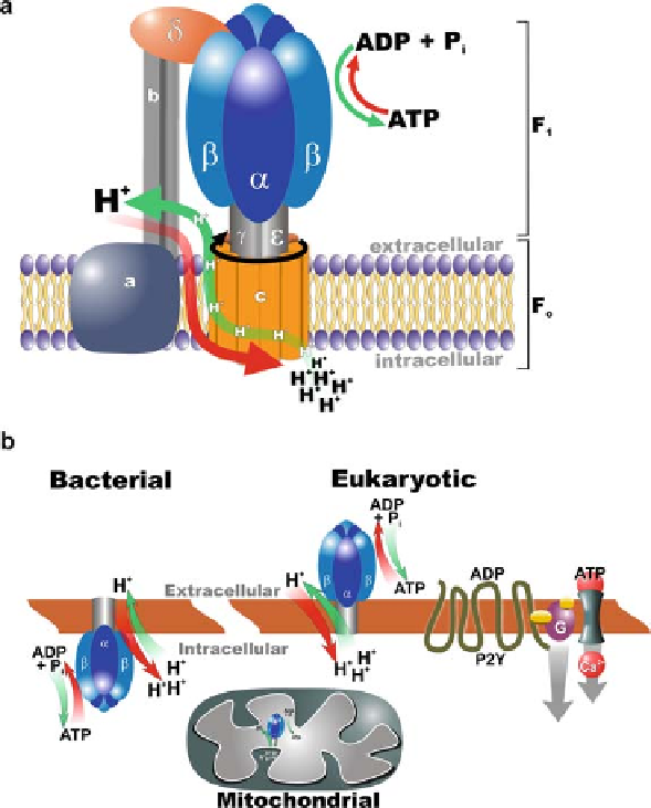
Fig. 9.1
Basic structure and cellular locations of F
1
F
0
ATP synthase. (
a
)F
1
F
0
ATP synthase con-
sists of a membrane-embedded component (F
0
) and a soluble portion (F
1
). Flow of protons down
their concentration gradient through F
0
causes rotation of the central stalk, leading to conforma-
tional changes of the
subunits and catalysis of ATP synthesis. The complex can also function in
reverse, coupling ATP hydrolysis with proton flow against the electrochemical gradient. (
b
)ATP
synthase is oriented in opposing directions in the plasma membrane of bacteria versus eukary-
otic cells. Cell surface ATP synthase in eukaryotes catalyzes ATP synthesis and hydrolysis in the
extracellular milieu. The resulting ATP can trigger cation influx into the cell through ATP-gated
ion channels (P2X purinoreceptors). ADP or ATP can bind to G-protein coupled receptors (P2Y
purinoreceptors) that can activate a variety of downstream signaling pathways. In mitochondria,
the electron transport chain generates a proton gradient across the inner mitochondrial membrane.
ATP synthase located within this membrane couples proton translocation with ATP synthesis in
the mitochondrial matrix. (Modified from Chi and Pizzo [14])
β
and high-density lipoprotein (HDL) endocytosis [24]. Other researchers have also
demonstrated colocalization of ATP synthase with caveolin-1 on the plasma mem-
brane of endothelial cells, supporting the theory proposed by Moser et al. that the
α
Search WWH ::

Custom Search