Biomedical Engineering Reference
In-Depth Information
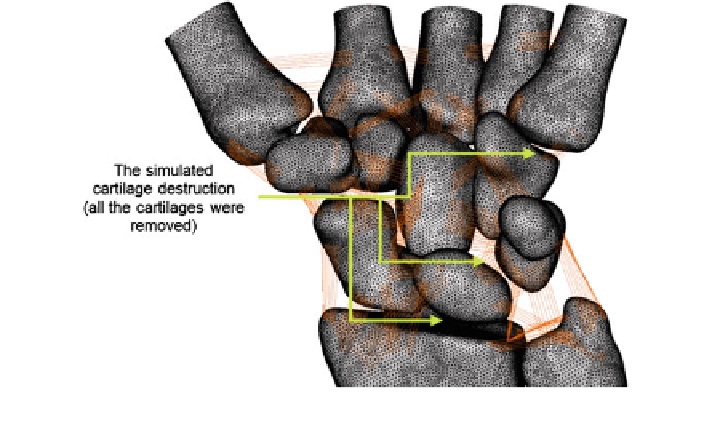
Fig. 5.1
Modelling of the cartilage destruction
1. Cartilage destruction [
1
-
3
].
2. Loss of carpal height due to bone destruction [
2
,
4
].
3. Dislocation of the carpus in the ulnar direction [
2
].
4. Dislocation of the proximal carpal row in the palmar and ulnar direction [
2
].
5. Scapholunar dissociation (SLD) with scapholunate advanced collapse wrist
arthritis (SLAC) stage 2 [
2
,
5
].
6. Dislocation of the scaphoid in the palmar direction due to the radial insertion
of the Testut ligament synovialitis [
2
].
7. Hand scoliosis due to ruptured tendon. This mechanism ends in a changed axis
of the wrist to the ulna with a consecutive rotation of the metacarpal bones in
the radial direction [
2
].
8. Reduction of contact between the lunate and radius [
2
].
9. Bone erosion [
2
,
3
,
6
-
8
].
10. Osteoporotic bone [
2
,
3
]. This criterion was simulated by reducing the elastic
modulus of the bones; 33 % for the cortical bone and 66 % for the cancellous
bone [
9
-
15
].
All these ten characteristics were utilised as a whole to construct the model of
the rheumatic wrist. The succeeding sections explained steps performed to simu-
late each characteristic.
5.1.1 Simulation of Cartilage Destruction
The cartilage destruction was modelled by removing all the articular cartilages to
simulate worst-case scenario (Fig.
5.1
). It was thus resulted in existence of gaps
Search WWH ::

Custom Search