Biomedical Engineering Reference
In-Depth Information
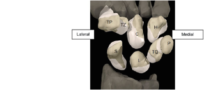
Fig. 1.3 Exploded
configuration for palmar view
of the wrist joint complex
showing different bone
structures of the eight carpal
bones. H hamate, C capitates,
TZ trapezoid, TP trapezium,
TQ triquetrum, P pisiform,
L lunate, S scaphoid [
8
,
9
]
As aforementioned, there are eight carpal bones where each of their names was
given according to their shape. Table
1.1
provides information on the structure for
each bone of the wrist joint.
1.3 Cartilage Structure
Cartilage comprises of a dense network of collagen fibers embedded in a gel-like
component of the ground substance namely chondroitin sulfate [
11
]. It lacks blood
vessels, lymphatics and nerves [
8
]. There are three types of cartilages: hyaline
cartilage, fibrocartilage and elastic cartilage. Articular cartilage which is composed
from hyaline cartilage is mainly located at the ends of long bones. It promotes
flexibility, support as well as smooth surfaces to assist movements of joints [
11
].
Four layers exist as shown in Fig.
1.4
with the topmost layer namely superficial
zone which has the greatest importance. Since articular cartilage functions as a
nearly frictionless bearing while uniformly transferring loads on underlying bone
preventing high stress concentrations [
12
], this layer acts by supporting more than
90 % of the compressive loads. This corresponding surface also functions to resist
shear forces generated by the joint movement. The fluid and collagen content in
the cartilage differ, which subsequently lower in the intermediate, deep and cal-
cified zones of articular cartilage. The collagen fibers determine its strength and its
resilience, which is an ability to restore its original shape after deformation
attributed to the presence of chondroitin sulfate.
There were numerous reports on works related to pathological condition of the
cartilage. One of them was a study on its poor capacity of repair and healing thus
resulted in cartilage degeneration [
13
]. This study has demonstrated that the onset
and progression of the disease was due to perturbation in underlying bone through
acute injuries [
14
]. It was expected that local alterations in the subchondral bone
Search WWH ::

Custom Search