Biomedical Engineering Reference
In-Depth Information
GPa in the transversal direction [585]. The elastic properties of bone
were successfully modeled at the level of mineralized collagen fibrils
via step-by-step homogenization from the staggered arrangement
of collagen molecules up to an array of parallel mineralized fibrils
[586]. Recent investigations revealed that bone deformation was not
homogeneous but distributed between a tensile deformation of the
fibrils and a shearing in the interfibrillar matrix between them [587,
588]. Readers, who are interested in further details, are addressed
to a good review on the effects of the microscopic and nano-scale
structure on bone fragility [589].
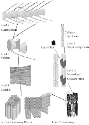
Figure 1.11
The seven hierarchical levels of organization of the zebrafish
skeleton bone. Level 1: Isolated crystals and part of a collagen
fibril with the triple helix structure. level 2: mineralized
collagen fibrils. level 3: The array of mineralized collagen
fibrils with a cross-striation periodicity of nearly 60-70 nm.
level 4: Two fibril array patterns of organization as found in
the zebrafish skeleton bone. level 5: The lamellar structure
in one vertebra. level 6: A vertebra. level 7: Skeleton bone.
reprinted from ref. [602] with permission. Other good
graphical sketches of the hierarchical structure of bones are
available in Refs. [544, 583, 584].


Search WWH ::

Custom Search