Biology Reference
In-Depth Information
FLUORESCENCE LABELING
3 x R1
3 x R2
3 x R3
3 x R4
Reference
state
(R)
Unlabeled
extracts
3 x R1
3 x R2
3 x R3
3 x R4
Internal
standard
(IS)
Cy2-labeled
extract mixture
IS = R(1-4) + A(1-4) + B(1-4) + C(1-4)
Unlabeled
extracts
A2
A3
A4
A1
B1
B2
B3
B4
C1
C2
C3
C4
Test
states
(A, B, C)
A1
A2
A3
A4
B1
B2
B3
B4
C1
C2
C3
C4
PREPARATION OF MIXTURES
R1
R2
R3
R4
R1
R2
R3
R4
R1
R2
R3
R4
+
+
+
+
+
+
+
+
+
+
+
+
IS
IS
IS
IS
IS
IS
IS
IS
IS
IS
IS
IS
+
+
+
+
+
+
+
+
+
+
+
+
A1
A2
A3
A4
B1
B2
B3
B4
C1
C2
C3
C4
2DE
B4
C4
A4
B3
C3
A3
B2
C2
A2
B1
C1
A1
IMAGE ACQUISITION (3-mode scanning)
Green
(532 nm)
Blue
(488 nm)
Red
(633 nm)
Cy3-image
Cy2-image
Cy5-image
IMAGE ANALYSIS (DeCyder)
Raw data
12 gels
= 36 gel images
DIA
intragel
comparison
1 gel
BVA
intergel
comparison
12 gels
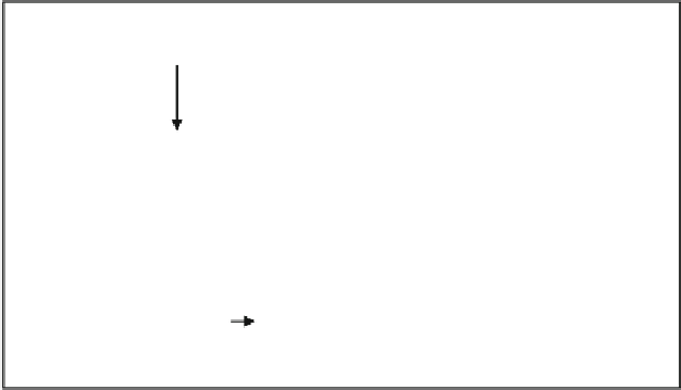
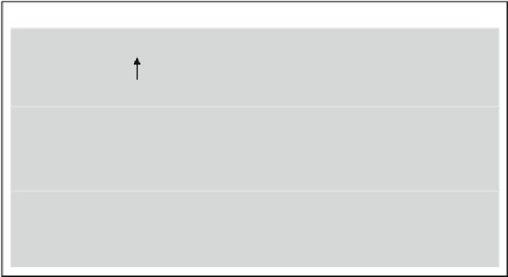
Fig. 3. Scheme of a typical 2D DIGE experiment consisting of fl uorescence labeling, preparation of mixtures of labeled
protein extracts, 2DE-based protein separation, image acquisition, and analysis (modifi ed from (
31
) ) .


Search WWH ::

Custom Search