Biology Reference
In-Depth Information
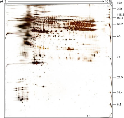
Fig. 2. Representative 2D image of ProteoMiner™ fractionated serum.
4. The PMT voltage for each channel is adjusted to ensure that
the maximum pixel values of the three channels only differ
within 5-10% and that the PMT voltages used do not saturate
all spots of interest in the 2D gel (see Note 14).
5. Two gels can be scanned simultaneously once the PMT voltages
are optimized. The images should be acquired at high resolu-
tion of 100 mm for image analysis.
6. The images fi les are kept with .gel extension.
7. After the gels are removed from the glass plates, they can be
placed in fi xing solution until use.
8. Scanned images are analyzed using the DeCyder™ 2D Differen-
tial Analysis Software version 6.5.
9. An example of a 2DE gel image for a ProteoMiner™-treated
serum is shown in Fig.
2
.
4. Notes
1. The ProteoMiner™ kit provides reagents enough for processing
ten reactions per samples. The lyophilized elution reagent after
reconstitution can only be stored up to 1 week at −20°C. It is
recommended to use freshly prepared buffer when conducting
Search WWH ::

Custom Search