Biology Reference
In-Depth Information
Internal
standard
Control
Treated
Cy5
Cy2
Cy3
Mix samples
2D-electrophoresis
Image Acquisition and Analysis
Protein ID
Fig. 1. Scheme of a DIGE experiment, using three fl uors and a single gel.
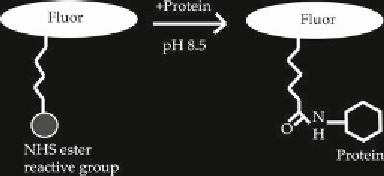
Fig. 2. Scheme of the DIGE labeling reaction.
method is referred to as “minimal labeling.” This ensures that the
stoichiometry of protein to fl uor, results in only 1-2% of the total
number of lysine residues being labeled.
After labeling, protein samples are mixed and separated on the
same 2D gel. Different protein extracts labeled with different


Search WWH ::

Custom Search