Agriculture Reference
In-Depth Information
compartmentalization is disrupted, resulting in leakage of calcium ions and hydrogen ions
from their storage compartments such as the cell wall and the vacuole. In addition, the
activities of key plasma membrane ATPases, such as the calcium and proton ATPase that
extrude the ions from the cytosol to the cell wall space, are negatively affected during
senescence or after ethylene treatment as demonstrated in carnation flower petals (Paliyath
and Thompson, 1988; Paliyath et al., 1997). Thus, reduced ATPase activity can also result
in a buildup of calcium ions and hydrogen ions in the cytoplasm. Such conditions lead to
the autocatalytic progression of membrane lipid degradation once it has been initiated by
hormones (ethylene and abscisic acid) or stress. Although there is a clear link between the
promotion of senescence by ethylene and enhanced membrane deterioration, the complete
sequence of signal transduction events involved in this link has not yet been established.
9.3.1 Changes in PLD activity during ripening
Previous studies have attempted to correlate increased membrane deterioration that occurs
during ripening and senescence to increased phospholipase D activity. Although such an
increase in PLD activity was noticeable in some senescing systems such as broccoli florets
(Deschene et al., 1991), it was not as distinct in systems such as carnation flower petals
(Paliyath et al., 1987) and tomato fruit (Jandus et al., 1997). Thus, increased phospholipid
degradation that occurs during ripening/senescence was linked to the activation of PLD by
factors such as increase in cytosolic calcium and a decrease in pH, membrane rigidification,
and fatty acid retailoring that increases the availability of preferred PLD substrates (Brown
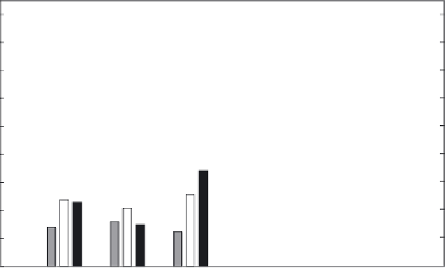
et al., 1990). We have examined PLD activity during fruit development using cherry toma-
toes, where the developmental stages are physiologically more precise and distinguishable.
PLD activity was determined in subcellular fractions comprising mitochondrial membranes,
microsomal membranes, and the cytosol (Fig. 9.5). Mitochondrial PLD activity remained
9
8
7
6
5
4
3
2
1
0
YNG
INT
MG
TOR
OR
RED
Developmental stages
Fig. 9.5
Changes in PLD activity during development of cherry tomato. PLD activity in mitochondrial (grey),
microsomal (unshaded), and cytosolic (dark) fractions was measured at young (YNG), intermediate (INT), mature
green (MG), turning orange (TOR), orange (OR), and red (RED) stages. (Reproduced with permission from
Pinhero et al., 2003.)


Search WWH ::

Custom Search