Biomedical Engineering Reference
In-Depth Information
thus resulting in a continuous film of the metal
oxides with embedded TMV, in analogy to the
films produced with ferritin.
The important observation after treating the
TMV was that the coating also occurred within
the hollow central channel of the virus. Trans-
mission electron microscopy (TEM) observa-
tions clearly showed an enhanced contrast in the
channel, which was not only proof of the pres-
ence of the metals but also that the structure of
the virus did not collapse.
The TMV is prone to break into smaller subu-
nits if mechanically treated. Such subunits were
also observed in the TEM after the ALD process.
Small subunits, dependent on their aspect ratio,
tend to lay upright, which confirmed that the
central channel of 4 nm in diameter was coated,
leaving behind a much narrower channel in the
center (
Figure 16.6
). This can be explained as fol-
lows: The precursors used for the ALD process
can access any available surface. In the case of the
TMV, the channel of 4 nm in diameter is acces-
sible to the precursors that attach and form the
metal oxide during the process. Once the deposit
in the central channel starts growing, the channel
opening shrinks. When it becomes too narrow for
the precursors to enter, the deposit will close the
opening and encapsulate the channel. The size of
the remaining channel should be a function of the
size of the molecular precursors.
The use of TMV as a template for ALD pro-
cessing enables additional functionalization.
This can be done by combining ALD with
approaches based on wet-chemical methods,
such as redox reactions or electroless deposition
[57-62]
. Also, adsorption of TMV on various
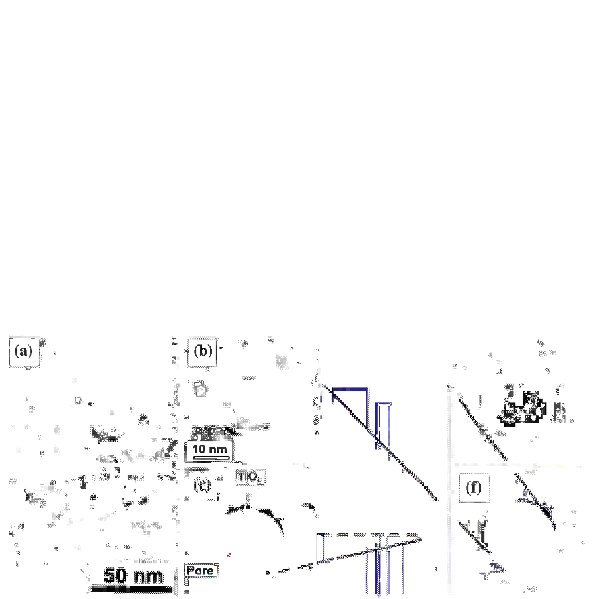
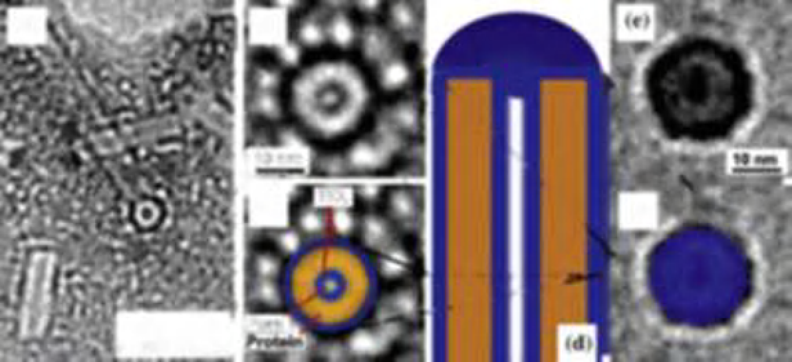
FIGURE 16.6
(a) TEM (200 kV) image of TMV treated with TiO
2
by ALD. A disk from a broken TMV (circular particle)
embedded in an amorphous TiO
2
film presents an axial view. The TiO
2
covering the interior channel appears hollow with a
channel diameter of 1-1.5 nm and a wall thickness of 1 nm. The covered inner channel of the virus appears brighter along
the axis, indicating a hollow TiO
2
nanotube. (b) Magnification of a further TiO
2
-covered disk showing a hollow area inside
the TiO
2
-coated interior channel of the virus. (c) Optically enhanced (b). The bright circle represents the viral protein sheath.
Darker circles show the TiO
2
coating of the viral surface (outer surface and channel surface). The surrounding gray area is
the embedding amorphous TiO
2
film. (d) Sketch of a cross-section of a TiO
2
-covered TMV. In the top part of the virus, no
channel is visible in the center; this part represents the assumed clogged area of the inner viral channel. (e) Magnification
of a further TiO
2
-covered disk showing a clogged interior channel of the virus. (f) Optically enhanced (e). Reprinted from
Ref.
29
. Copyright © 2006, with permission from the American Chemical Society.