Biology Reference
In-Depth Information
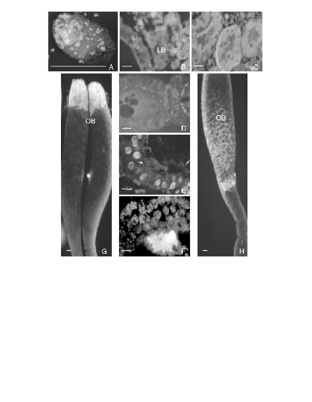
FIGURE 5.1
(Color Ýgure follows p. 206.)
Fluorescent
in situ
hybridization (FISH) of
Sitophilus
oryzae
intracellular bacteria. SpeciÝc oligonucleotide probes were designed by sequence alignment of
Wolbachia
and
SOPE 16S rDNA. Two
Wolbachia
probes (W1, W2) 5
end labeled with rhodamine were used to increase the
signals. The SOPE probe (S) was 5
end labeled with rhodamine except in panel B (with Þuorescein).
Hybridization was performed as described by Heddi et al. (1999). Slides were mounted in Vectashield medium
containing DAPI. (A) Bacteriocyte labeled with W1W2. (B) Larval bacteriome (LB) labeled with W1W2 and
S (Þuorescein). (C, D, E, F, and H) Adult mesenteric caeca bacteriomes, oocyte, follicular cells, testis, and
ovary, respectively, labeled with W1W2. (G) Ovary labeled with S. Scale bar = 10 ´m.
designed for SOPE and
Wolbachia
16S rDNA showed that although SOPE was limited to the
were disseminated throughout all insect tissues even within the bac-
teriocytes, coexisting with SOPE (Figure 5.1).
bacteriocytes,
Wolbachia
spp. are present in low density in
muscles, adipocytes, and the intestine, whereas they are highly abundant in the male and female
germ cells, located around the periplasmic membrane of the oocyte and mixed with many
spermatid nuclei in the testis (Figure 5.1). Here also
Wolbachia
do not interact with the host
in the same way as integrated endosymbionts do because they are not exclusive to speciÝc host
cells (such as the bacteriocytes), but rather germ cells are the privileged site for the development
of these bacterial cells.
Wolbachia