Biology Reference
In-Depth Information
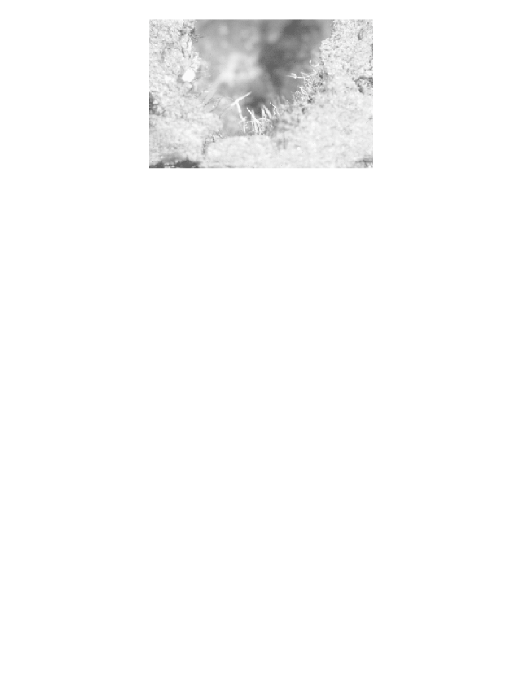
FIGURE 7.3
(Color Ýgure follows p. 206.)
Conidiophores (anamorph) of
Ophiostoma clavigerum
lining a
pupal chamber of
Dendroctonus ponderosae
.
probably important for some fungi in long-distance dispersal between host trees (Malloch and
Blackwell, 1993). The ascospores produced by
are coated with
an adhesive material and are of various shapes that allow for multiple contact points with the vector,
ensuring that they are not easily removed in transit. The adhesive coats of some
Ophiostoma
and
Ceratocystiopsis
Ophiostoma
ascospores disperse in resin but not in water, which may provide a mechanism for removal of the
sticky spores only when an appropriate substrate (i.e., the new host tree) is encountered (Whitney
and Blauel, 1972).
The ophiostomatoid fungi also produce a wide range of anamorphs that produce conidia (asexual
spores) in slimy masses that readily adhere to the insect cuticle (Tsuneda and Hiratsuka, 1984;
Tsuneda, 1988; Malloch and Blackwell, 1993). The conidia are often round, cylindrical, or oval,
permitting only one contact point with the vector arthropod, which may allow the conidia to be
easily dislodged and dispersed within beetle galleries (Malloch and Blackwell, 1993). The conidia
are also often found in pits on the beetle exoskeleton or in mycangia, indicating they also play a
critical role in long-distance dispersal for some species.
The basidiomycetes (
spp.) associated with bark beetles produce both chlamy-
dospores and conidia. However, in mycangia, only chlamydospores or a yeast-like form of the
fungus is present (Barras and Perry, 1972; Happ et al., 1976; Goldhammer et al., 1990).
For beetles possessing sac mycangia, apparently only the anamorph is acquired and dissemi-
nated in the structure, although sexual reproduction of the fungi may also occur (Paine and Birch,
1983; Moser et al., 1995). For these beetles often only the anamorph is produced in the pupal
chambers (Figure 7.3) where mycangia of teneral adults are charged with fungal propagules prior
to dispersal (Whitney, 1971). In contrast, ascomata often form in old galleries distant to where
teneral adult beetles develop (
Figure 7.4)
,
and therefore contact between the sexual stage and new
adults prior to emergence is unlikely. If and how the sexual stages of these fungi are disseminated
has posed a difÝcult problem for investigators. The answer to this conundrum for at least one bark
beetle system may be found by looking at phoretic mites associated with the insect (Klepzig et al.,
2001). The ascospores of a mycangial fungus of
Entomocorticium
are transported in
sporothecae of phoretic mites while the conidia are carried in the mycangium of the host beetle
(Moser et al., 1995). The mites colonize the insect host within the pupal chambers prior to beetle
emergence and dispersal. It is not known if mites are important vectors of ascospores of mycangial
fungi in other bark-beetle systems.
For other fungi associated with sac mycangia, the production of the sexual state may be rare
or even lacking. The sibling species,
D. frontalis
,
C. ranaculosus,
in
sac mycangia. Laboratory pairings of this fungus have not produced ascomata, and there is some
question as to whether a sexual state exists for this fungus (D.L. Six, T.C. Harrington, D. McNew,
D. ponderosae
and
D. jeffreyi
, both carry
O. clavigerum