Biomedical Engineering Reference
In-Depth Information
It is certainly one of the strengths of optical microscopy to be able to image
samples in water-based solution. This way, live cells or tissues can be imaged. Fluo-
rescence microscopy enables to observe only the objects carrying a fluorescent dye
(Figure 8.12). The proteins of interest are then localized by comparing images in
fluorescence mode to images in white light mode. By using two dyes and working
with two colors, two different objects can be colocalized.
Many solutions exist that can increase the contrasts of a particular image de-
pending on the characteristics of the sample : The polarization of the light, its angle
of incidence, the interference between two light rays are strategies used alone or in
conjunction and that are commercially available [24].
The confocal microscope [25] works on a somewhat different principle. The
image is formed point after point. Here, the light source is a point source. The point
image in the sample emits light that is collected by a detector through a pinhole,
confocal with the source. This way, only the light that one wants to collect is indeed
collected; the light emitted by other parts of the sample whose fluorescence is also
excited by the illumination (the parts of the sample in the cone of light produced by
the microscope objective) is largely excluded from the detector (Figure 8.13). The
sample is then scanned in order to make the full scale image. Why is this interesting?
In classical microcopy, all the fluorescence light is collected, and although there is a
maximum of intensity at the focal point, the image is blurred by this background.
The confocal technique is very efficient in rejecting unwanted out-of-focus light.
“Slices” at a given height are performed by scanning the sample in the
x-y
plane and
three-dimensional images can then be reconstructed by combining these slices. As
it is a scanning technique, it is somewhat slow and thus not very efficient to study
fast dynamics.
A further refinement of confocal microscopy is the two-photon microscopy [25]
that uses a nonlinear effect to excite fluorescence only at the focal point leaving un-
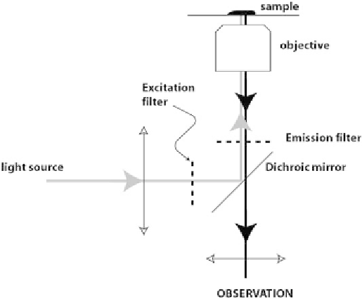
Figure 8.12
Principle of fluorescence microcopy. In this particular configuration, the excitation
light and the fluorescence emission go trough the objective (epifluorescence).