Biomedical Engineering Reference
In-Depth Information
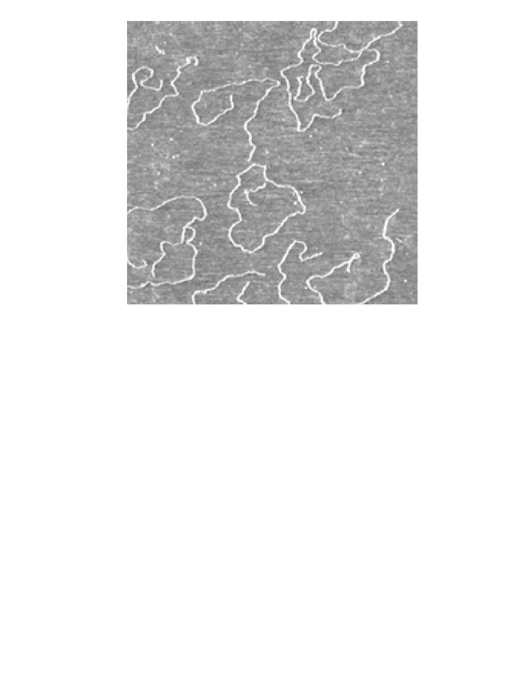
Figure 8.3
Atomic force microscopy of DNA molecules adsorbed on a solid surface (see a descrip-
tion of the technique in Section 8.3.1.3). The image is color-coded in height so that the height
difference between the white color and the dark color is ~ 1 nm. The size of the image is 1.5
m
m.
From such images the contour length of these molecules can be accurately measured as well as the
localization of potentially interacting proteins. (From [3].)
structures. The shape imposed on these molecules by this folding plays an impor-
tant role in the translation into proteins.
The folding of these molecules is fixed by their sequence, hence by the DNA
sequence. Their structures correspond to a minimum of energy and can now be ac-
curately computed for reasonably long molecules (up to a few 1,000 nucleotides)
[6] (Figure 8.4).
Because it is an indicator of gene expression, mRNA has a central position in
functional genomics and mRNA is one of the major targets of the DNA arrays de-
scribed above. By hybridization with the small DNA sequences spotted on the array,
the levels of particular RNAs are measured and, from there, one gets some infor-
mation on the amount of the translated functional proteins. Practically, it is much
easier to work with RNA than with proteins, and since their levels are strongly
correlated, it is quite useful information.
8.1.1.3 Proteins
Proteins are the product of the translation of RNA by ribosomes in the cytoplasm.
Along the RNA strand, three consecutive nucleotides, a codon, are translated into
an amino acid in a very robust way following the so-called genetic code.