Biomedical Engineering Reference
In-Depth Information
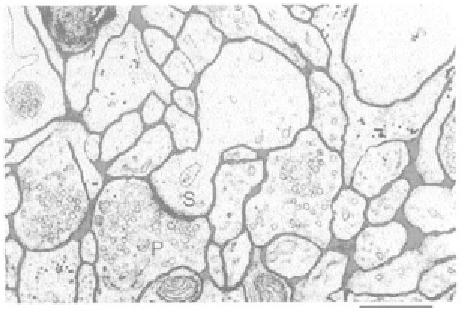
Figure 5.30
Geometry of extracellular space from [15]—an electromicrograph of a small region of
rat cortex. The ECS is in dark on the picture. “Lakes” or intercleft spaces can be seen at the bottom
right where the extracellular space widens.
In the biological field, the delivery of drugs in the human body, especially in
clusters of cells, is an utmost important problem requiring the knowledge of dif-
fusion in a complex, confined geometry. In this case, there are two different types
of media: the extracellular space (ECS) and the cells; these two types are separated
by the cell membrane. Diffusion of biological molecules first takes place inside the
extracellular space. After the molecules have penetrated the cells through the cell
membranes (which is called uptake), the molecules diffuse inside the cells. In this
section, we focus on the diffusion inside the ECS.
Because cellular uptake is not immediate, the biological cluster of cells may be
seen as porous media, where the cells are the “solid grains” and the extracellular
space (ECS) the “pores” (Figures 5.30 and 5.31). In the particular case of tumoral
cells, the extracellular path is called the tumor interstitial matrix (IM) [14].
Figure 5.31
Cell arrangement in the human skin from [16]. The shape of the cells is regular, but the
anisotropy of the ECS changes from top to bottom. The typical width of the ECS is a few microns.